This site needs JavaScript to work properly. Please enable it to take advantage of the complete set of features!
Skip to main page content
U.S. flag

An official website of the United States government

Dot gov

The .gov means it’s official.
Federal government websites often end in .gov or .mil. Before sharing sensitive information, make sure you’re on a federal government site.

Https

The site is secure.
The https:// ensures that you are connecting to the official website and that any information you provide is encrypted and transmitted securely.

NIH NLM Logo
Log in
Access keys NCBI Homepage MyNCBI Homepage Main Content Main Navigation
. 2014 Mar;16(3):396-410.
doi: 10.1111/cmi.12224. Epub 2013 Nov 3.

Identification of a type IV secretion substrate of Brucella abortus that participates in the early stages of intracellular survival

Affiliations

Identification of a type IV secretion substrate of Brucella abortus that participates in the early stages of intracellular survival

Peter H Döhmer et al. Cell Microbiol. 2014 Mar.

Abstract

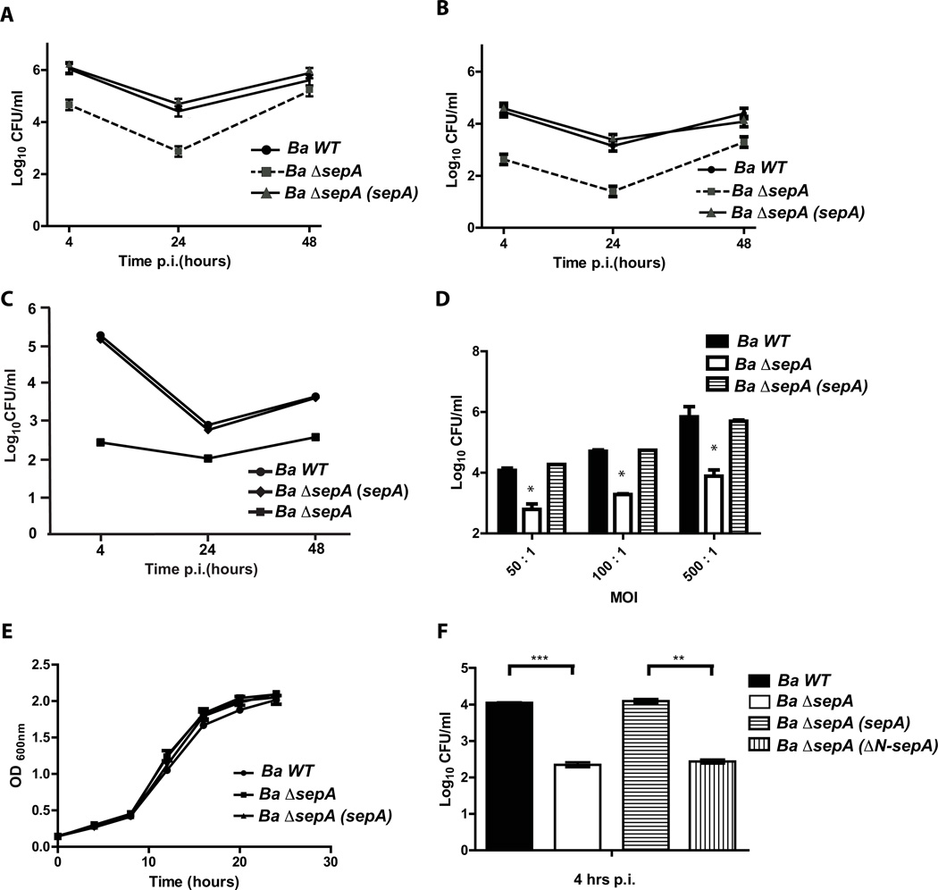
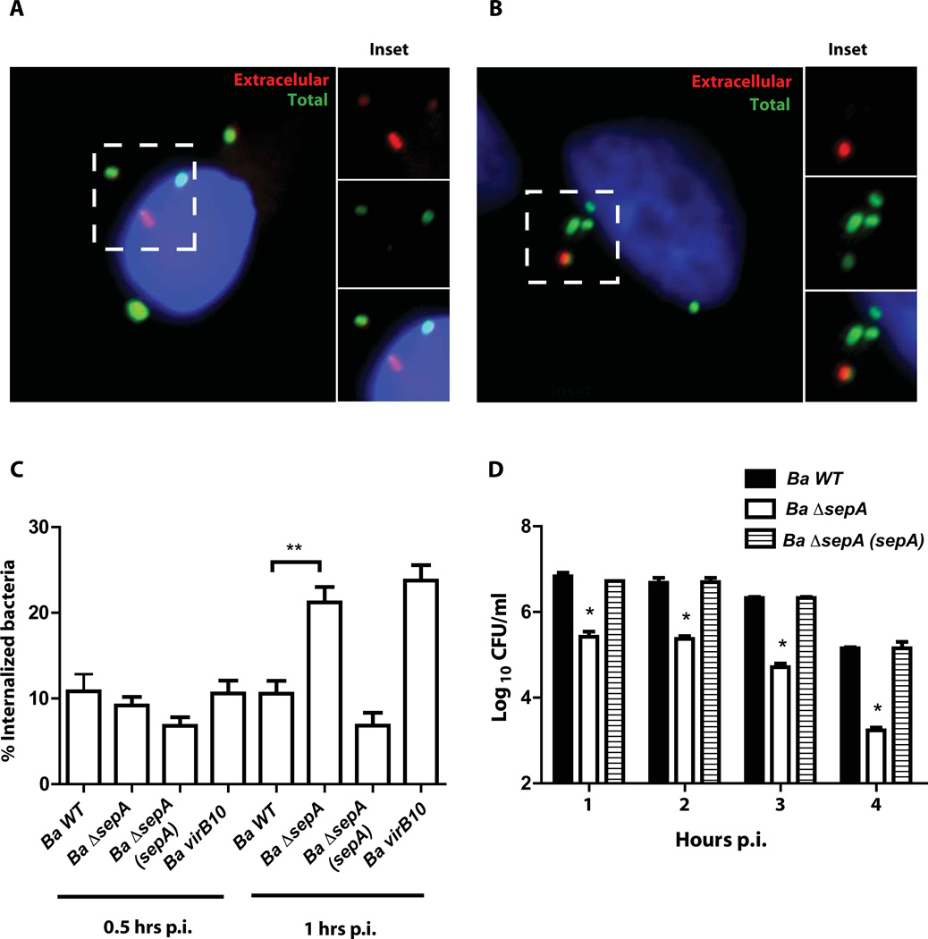
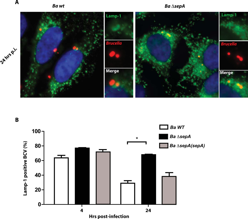
Brucella abortus, the aetiological agent of bovine brucellosis, is an intracellular pathogen whose virulence is completely dependent on a type IV secretion system. This secretion system translocates effector proteins into the host cell to modulate the intracellular fate of the bacterium in order to establish a secure niche were it actively replicates. Although much has been done in understanding how this secretion system participates in the virulence process, few effector proteins have been identified to date. We describe here the identification of a type IV secretion substrate (SepA) that is only present in Brucella spp. and has no detectable homology to known proteins. This protein is secreted in a virB-dependent manner in a two-step process involving a periplasmic intermediate and secretion is necessary for its function. The deletion mutant showed a defect in the early stages of intracellular replication in professional and non-professional phagocytes although it invades the cells more efficiently than the wild-type parental strain. Our results indicate that, even though the mutant was more invasive, it had a defect in excluding the lysosomal marker Lamp-1 and was inactivated more efficiently during the early phases of the intracellular life cycle.


Conflict of interest statement

The authors declare no conflict of interest.

Figures

Figure 1
Figure 1. The protein product of Bab1_1492 is secreted during the infection of host cells
A. Schematic representation of the screening method used to identify novel secreted virulence effectors in Brucella abortus. B. Immunofluorescence of J774 A.1 cells infected with a B. abortus strain coding for Bab1_1492-3xFLAG in a replicative vector at 4 hrs post-infection. Red, Brucella; green, Flag. C. Immunofluorescence of J774 A.1 cells infected with a B. abortus strain coding for Bab1_0740-3xFLAG in a replicative vector at 4 hrs post-infection. Red, Brucella; green, Flag. D. Western-blot with an anti-FLAG of the strains shown in panels B and C.
Figure 2
Figure 2. Confocal analysis of the secretion of the protein product of Bab1_1492
Confocal images of J774 A.1 cells infected with the B. abortus strains coding for Bab1_1492-3xFLAG (A) or Bpe123-3xFLAG (B) in replicative vectors at 4 hrs post-infection. C. A 45° rotation view of the line traced in the inset of panel A. D. Quantification of the red and green fluorescence intensity over the line traced in the inset of panel A showing a correlation between both markers. Red, Brucella; green, FLAG.
Figure 3
Figure 3. Secretion of SepA is a virB dependent process
A. Representative Immunofluorescence of J774 A.1 cells infected with B. abortus wild type, virB10 and ΔvirB11 mutant strains coding for SepA-3xFLAG in a replicative vector at 4 hrs post-infection. Red, Brucella; green, FLAG. B. Quantification of the percent of SepA secreting bacteria of the representative images shown in A. *P<0.05. C. Western-blot with an anti-FLAG of the strains showing equivalent levels of expression of SepA.
Figure 4
Figure 4. Secretion of SepA involves a periplasmic intermediate
A. Western-blot with an anti-FLAG, anti-GroEL and anti OMP1 monoclonal antibodies of periplasmic or cytoplasmic fractions of a periplasmic fractionation assay performed with B. abortus chromosomal Bab2_0448-3xFLAG tagged strain carrying the plasmid coding for SepA-3xFLAG. Schematic representation of the ΔN-SepA-3xFLAG (ΔN-SepA) construction carrying a N-terminal 25 amino acid deletion. B. Immunofluorescence of J774 A.1 cells infected with B. abortus strains carrying plasmids coding for SepA-3xFLAG (SepA) or ΔN-SepA-3xFLAG (ΔN-SepA) at 4 hrs post-infection. Red, Brucella; green, FLAG. C. Western-blot showing the expression levels of SepA and ΔN-SepA in Wild-type B. abortus. Loading control, OMP1. D. Western-blot with an anti-FLAG, anti-GroEL and anti OMP1 monoclonal antibodies of periplasmic or cytoplasmic fractions of a periplasmic fractionation assay performed with B. abortus virB10 and virB11 mutant strains carrying the plasmid coding for SepA-3xFLAG.
Figure 5
Figure 5. SepA participates in the early stages of intracellular replication
A. Intracellular replication of B. abortus wild type and ΔsepA deletion strains in HeLa cells determined by the gentamicin protection assay. B. Intracellular replication of B. abortus wild type and ΔsepA deletion strains in J774 A.1 cells determined by the gentamicin protection assay. C. Intracellular replication of B. abortus wild type and ΔsepA deletion strains in bone marrow derived macrophages determined by the gentamicin protection assay D. Viable bacteria of the wild type (Ba wt), Ba ΔsepA mutant and complemented strains at 4 hrs post-infection measured by an antibiotic protection assay in J774 A.1 cells with different multiplicity of infection ratios (50, 100 and 500). E. Growth curve of the wild type and the ΔsepA deletion strain in TSB. F. Complementation of the 4 hrs phenotype with the construction lacking the N-terminal periplasmic localization signal. **P<0.002 and ***P<0.001.
Figure 6
Figure 6. The ΔsepA mutant strain invades cells more efficiently
A and B. In/out staining of the wild type strain (Ba wt) and the ΔsepA (Ba ΔsepA) mutant strain respectively at 1 hr post-infection in J774 A.1 cells. C. Quantification of the immunofluorescence shown in panels A and B. Ba virB10, assay performed with the B. abortus virB10 mutant. **P<0.002. D. Intracellular viable bacteria of the wild type mutant and complemented strains at 1, 2, 3 and 4 hrs post-infection determined by an antibiotic protection assay. *P<0.001.
Figure 7
Figure 7. The ΔsepA mutant strain is affected in its capacity to exclude the lysosomal marker Lamp-1
A. Representative immunofluorescence images of Lamp-1 positive and negative BCVs of the wild type (Ba wt) and mutant (Ba ΔsepA) strains in infected HeLa cells at 24 hours post-infection. Red, Brucella; green, Lamp-1. B. Quantification of the percent of Lamp-1 positive BCVs at 4 and 24 hrs post-infection with the wild type and the ΔsepA mutant strains.
Figure 8
Figure 8. Over-expression of SepA affects virulence in mice
A and B. Spleen colonization at 15 days post-infection of mice intraperiteonally infected with the wild type (Ba wt), sepA mutant (Ba ΔsepA), complemented (Ba ΔsepA(sepA)) or SepA over-expressing (Ba wt(sepA)) strains. Bacterial load was determined as indicated in Experimental procedures. *P<0.002. C. Western-blot with a monoclonal anti-O-antigen antibody (M84) against whole bacteria showing that neither the mutant nor the over-expressing strains are rough.

References

    1. Aderem A, Underhill DM. Mechanisms of phagocytosis in macrophages. Annu Rev Immunol. 1999;17:593–623. - PubMed
    1. Alvarez-Martinez CE, Christie PJ. Biological diversity of prokaryotic type IV secretion systems. Microbiol Mol Biol Rev. 2009;73:775–808. - PMC - PubMed
    1. Arocena GM, Sieira R, Comerci DJ, Ugalde RA. Identification of the quorum-sensing target DNA sequence and N-Acyl homoserine lactone responsiveness of the Brucella abortus virB promoter. J Bacteriol. 2010;192:3434–3440. - PMC - PubMed
    1. Arocena GM, Zorreguieta A, Sieira R. Expression of VjbR under nutrient limitation conditions is regulated at the post-transcriptional level by specific acidic pH values and urocanic acid. PLoS One. 2012;7:e35394. - PMC - PubMed
    1. Atluri VL, Xavier MN, de Jong MF, den Hartigh AB, Tsolis RE. Interactions of the human pathogenic Brucella species with their hosts. Annu Rev Microbiol. 2011;65:523–541. - PubMed

Publication types

LinkOut - more resources

Cite

AltStyle によって変換されたページ (->オリジナル) /