This site needs JavaScript to work properly. Please enable it to take advantage of the complete set of features!
Skip to main page content
U.S. flag

An official website of the United States government

Dot gov

The .gov means it’s official.
Federal government websites often end in .gov or .mil. Before sharing sensitive information, make sure you’re on a federal government site.

Https

The site is secure.
The https:// ensures that you are connecting to the official website and that any information you provide is encrypted and transmitted securely.

NIH NLM Logo
Log in
Access keys NCBI Homepage MyNCBI Homepage Main Content Main Navigation
. 2013 Sep;52(5):545-52.

Immunopathologic characterization of naturally acquired Trypanosoma cruzi infection and cardiac sequalae in cynomolgus macaques (Macaca fascicularis)

Affiliations

Immunopathologic characterization of naturally acquired Trypanosoma cruzi infection and cardiac sequalae in cynomolgus macaques (Macaca fascicularis)

Harshan Pisharath et al. J Am Assoc Lab Anim Sci. 2013 Sep.

Abstract

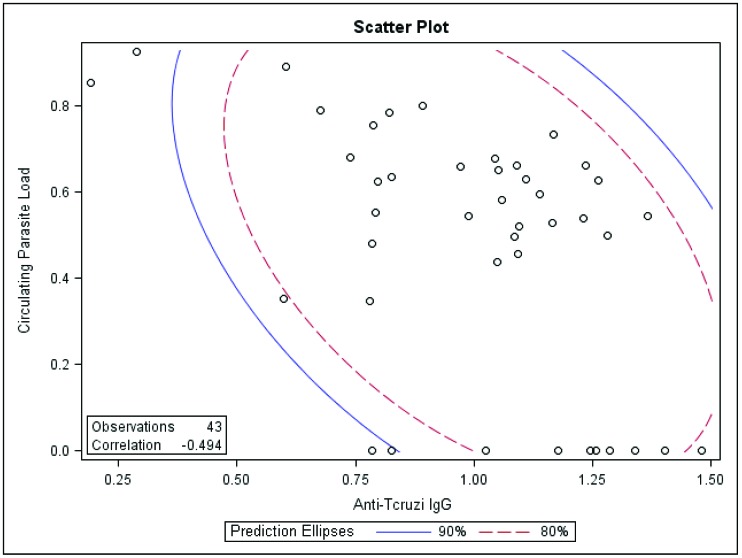
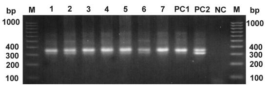
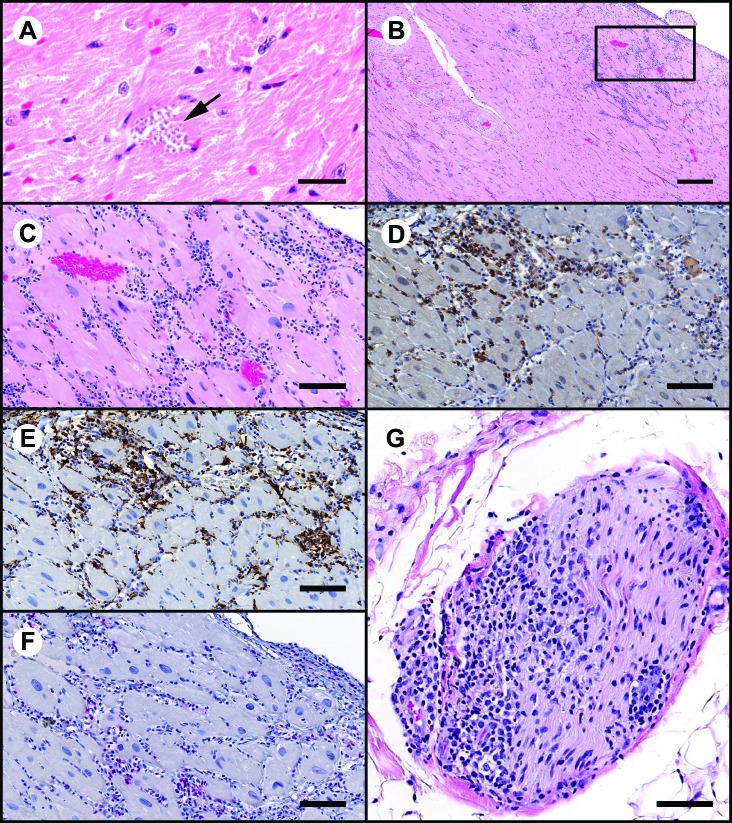
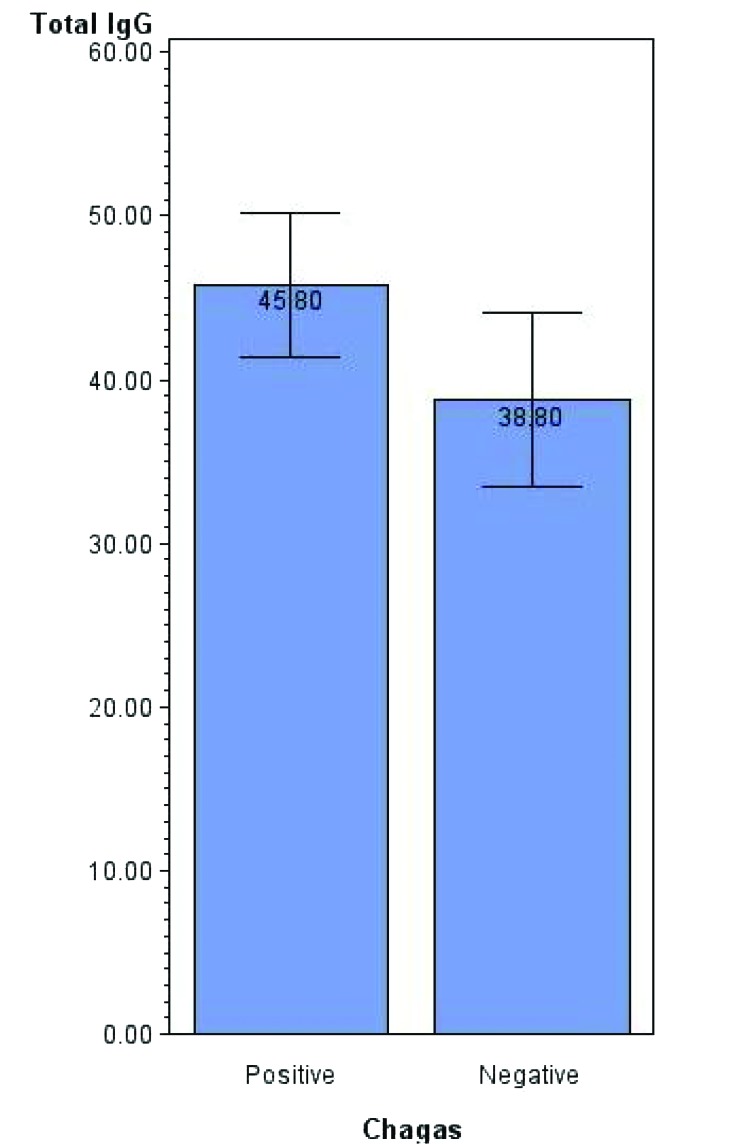
Trypanosoma cruzi, the causative agent of Chagas disease, is endemic in south Texas due to the abundant vector and wild small mammalian reservoir populations. This situation predisposes nonhuman primate colonies exposed to outdoor housing to infection from ingestion or bite of triatomid insects. Using a T. cruzi-specific real-time PCR and Trypanosome spp.-specific ELISA, we revealed a prevalence rate of 8.5% in a colony of outdoor-housed cynomolgus macaques. By using a discriminating kinetoplastid minicircle PCR, we eliminated the possibility of mixed prevalence with nonpathogenic trypanosomes and showed the ELISA results were specific for T. cruzi. In this study, we found an inverse relationship between antibody titers and circulating parasite load. Also, 23% of T. cruzi IgG ELISA-positive macaques were negative by real-time PCR. Furthermore, in a subset of infected macaques, cardiac tissue was infiltrated by inflammatory mononuclear cells and contained T. cruzi genomic and kinetoplast DNA despite lacking microscopic evidence of discrete parasite stages. In addition, 19% of the infected macaques had titers for cardiac troponin I autoantibody, which could contribute to autoimmune myocarditis or interfere with circulating troponin I measurements. These findings indicate the possibility of T. cruzi to interfere with the assessment of cardiac safety signals in preclinical toxicology and safety pharmacology studies and the necessity for prestudy screening for T. cruzi in outdoor-housed nonhuman primates from endemic areas.


Figures

Figure 1.
Figure 1.
Correlation (P = 0.002) between circulating parasite load (expressed as the ratio of the log of absolute copy number of T. cruzi nuclear satellite DNA to that of GAPDH) with antiT. cruzi IgG titer expressed in OD units.
Figure 2.
Figure 2.
Kinetoplast minicircle DNA detection by PCR. A 300- or 330-bp fragment (or both) amplified from T. cruzi minicircle DNA was present in all tested samples. M, DNA ladder marker (bp); lanes 1 through 7, pooled DNA pool samples (each pool contained 4 or 5 individual DNA samples that had been previously tested positive in T. cruzi real-time PCR); positive controls showing 330 bp (PC1) and 300- and 330-bp bands (PC2); nc, negative control (no-template control).
Figure 3.
Figure 3.
Sections of myocardium demonstrating areas of inflammation and characterization of the inflammatory cell infiltrates by IHC. (A) Cluster of T. cruzi amastigotes (arrow). Hematoxylin and eosin stain; bar, 20 μm. (B) Low-power magnification demonstrating moderate to marked subepicardial inflammatory cell infiltrates. Hematoxylin and eosin stain; bar 300μm. The boxed area is shown below. (C) High-power magnification from section in panel B. Inflammatory cell infiltrates are primarily mononuclear cells with fewer neutrophils. Hematoxylin and eosin stain; bar, 100 μm. (D and E) IHC peroxidase procedure with diaminobenzidine chromagen (brown). A considerable proportion of the mononuclear cell infiltrates are positive for (D) CD8 (lymphoid origin) and (E) CD68 (monocyte–macrophage origin). Bar, 100 μm. (F) IHC alkaline phosphatase procedure with Vector red chromagen (red). A small proportion of inflammatory cells are CD4+. Bar, 100 μm. G) Section of nerve at the heart base that is infiltrated by inflammatory cells. Hematoxylin and eosin stain; bar, 80 μm.
Figure 4.
Figure 4.
Comparison (mean ± SE, expressed as mg/mL; P = 0.041) of total circulating IgG levels in T. cruzi-infected and uninfected groups.

References

    1. Andrade MC, Dick EJ, Jr, Guardado-Mendoza R, Hohmann ML, Mejido DC, VandeBerg JL, DiCarlo CD, Hubbard GB. 2009. Nonspecific lymphocytic myocarditis in baboons is associated with Trypanosoma cruzi infection. Am J Trop Med Hyg 81:235–239 - PMC - PubMed
    1. Avila JL. 1992. Molecular mimicry between Trypanosoma cruzi and host nervous tissues. Acta Cient Venez 43:330–340 - PubMed
    1. Bermejo DA, Amezcua Vesely MC, Khan M, Acosta Rodriguez EV, Montes CL, Merino MC, Toellner KM, Mohr E, Taylor D, Cunningham AF, Gruppi A. 2011. Trypanosoma cruzi infection induces a massive extrafollicular and follicular splenic B-cell response which is a high source of nonparasite-specific antibodies. Immunology 132:123–133 - PMC - PubMed
    1. Bern C, Kjos S, Yabsley MJ, Montgomery SP. 2011. Trypanosoma cruzi and Chagas disease in the United States. Clin Microbiol Rev 24:655–681 - PMC - PubMed
    1. Bonney KM, Taylor JM, Daniels MD, Epting CL, Engman DM. 2011. Heat-killed Trypanosoma cruzi induces acute cardiac damage and polyantigenic autoimmunity. PLoS ONE 6:e14571. - PMC - PubMed

MeSH terms

Cite

AltStyle によって変換されたページ (->オリジナル) /