This site needs JavaScript to work properly. Please enable it to take advantage of the complete set of features!
Skip to main page content
U.S. flag

An official website of the United States government

Dot gov

The .gov means it’s official.
Federal government websites often end in .gov or .mil. Before sharing sensitive information, make sure you’re on a federal government site.

Https

The site is secure.
The https:// ensures that you are connecting to the official website and that any information you provide is encrypted and transmitted securely.

NIH NLM Logo
Log in
Access keys NCBI Homepage MyNCBI Homepage Main Content Main Navigation
Case Reports
. 2013 Oct;32(10):1341-5.
doi: 10.1007/s10096-013-1885-x. Epub 2013 May 14.

Another case of "European hantavirus pulmonary syndrome" with severe lung, prior to kidney, involvement, and diagnosed by viral inclusions in lung macrophages

Affiliations
Case Reports

Another case of "European hantavirus pulmonary syndrome" with severe lung, prior to kidney, involvement, and diagnosed by viral inclusions in lung macrophages

M Gizzi et al. Eur J Clin Microbiol Infect Dis. 2013 Oct.

Abstract

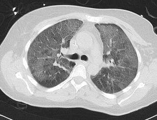
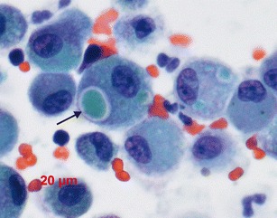
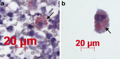
Puumala virus (PUUV) is considered a classic Old World etiologic agent of nephropathia epidemica (NE), or hemorrhagic fever with renal syndrome (HFRS). HFRS is considered to be distinct from hantavirus (cardio-)pulmonary syndrome (HPS or HCPS), described in the New World. Here, we report a severe case, which fulfilled most, if not all, Centers for Disease Control and Prevention (CDC) criteria for HPS, needing non-invasive ventilation and subsequent acute hemodialysis. However, the etiological agent was PUUV, as proved by serological testing, real-time polymerase chain reaction (PCR), and sequencing. Viral antigen was detected by specific anti-PUUV immunostaining, showing, for the first time, greenish intracytoplasmic inclusions in bronchoalveolar lavage (BAL) macrophages. This case definitely confirms that HPS can be encountered during PUUV infections. Interestingly, special findings could render the diagnosis easier, such as greenish homogeneous cytoplasmic inclusions, surrounded by a fine clear halo in BAL macrophages. Therefore, although the diagnosis remains difficult before the onset of renal involvement, the occurrence of severe respiratory failure mimicking community-acquired pneumonia must alert the clinician for possible HPS, especially in endemic areas.


Figures

Fig. 1
Fig. 1
Chest X-ray at hospital admission [day 5 post onset of symptoms (POS)] showing bilateral pulmonary interstitial infiltrates
Fig. 2
Fig. 2
Chest computed tomography (CT) scan (day 8 POS) showing bilateral ground-glass pattern
Fig. 3
Fig. 3
Bronchoalveolar lavage (BAL) macrophages containing greenish cytoplasmic inclusions surrounded by a clear halo (arrow, Papanicolaou stain, magnification ×ばつ40)
Fig. 4
Fig. 4
a, b Macrophage inclusions stained positively with specific Puumala virus (PUUV) antibody [arrow, magnification ×ばつ60 (a), ×ばつ100 (b)]
Fig. 5
Fig. 5
Phylogenetic tree based on Belgium and European PUUV S segment sequences. Sin Nombre virus (SNV), the typical American etiologic agent of the hantavirus pulmonary syndrome (HPS), is used as an outgroup in the analysis. The patient lineage is marked by a black square, and is referred to as "Doische", the putative place of infection in the densely forested South of Belgium, near the French border. For the Belgian strains, PUUV isolates from different regions are used. The evolutionary history was inferred using the neighbor-joining method. The bootstrap consensus tree inferred from 1,000 replicates is taken to represent the evolutionary history of the taxa analyzed. Bootstrap values less than 50 % are not shown. GenBank accession numbers are shown in italics. The scale bar represents the number of base substitutions per site

References

    1. Jonsson CB, Figueiredo LTM, Vapalahti O. A global perspective on hantavirus ecology, epidemiology, and disease. Clin Microbiol Rev. 2010;23:412–441. doi: 10.1128/CMR.00062-09. - DOI - PMC - PubMed
    1. Hjelle B, Torres-Pérez F. Hantaviruses in the Americas and their role as emerging pathogens. Viruses. 2010;2:2559–2586. doi: 10.3390/v2122559. - DOI - PMC - PubMed
    1. Clement J, Maes P, Lagrou K, van Ranst M, Lameire N. A unifying hypothesis and a single name for a complex globally emerging infection: hantavirus disease. Eur J Clin Microbiol Infect Dis. 2012;31:1–5. doi: 10.1007/s10096-011-1456-y. - DOI - PMC - PubMed
    1. Duchin JS, Koster FT, Peters CJ, et al. Hantavirus pulmonary syndrome: a clinical description of 17 patients with a newly recognized disease. The Hantavirus Study Group. N Engl J Med. 1994;330:949–955. doi: 10.1056/NEJM199404073301401. - DOI - PubMed
    1. Clement J, Colson P, McKenna P. Hantavirus pulmonary syndrome in New England and Europe. N Engl J Med. 1994;331:545–546. doi: 10.1056/NEJM199408253310813. - DOI - PubMed

Publication types

MeSH terms

LinkOut - more resources

Cite

AltStyle によって変換されたページ (->オリジナル) /