This site needs JavaScript to work properly. Please enable it to take advantage of the complete set of features!
Skip to main page content
U.S. flag

An official website of the United States government

Dot gov

The .gov means it’s official.
Federal government websites often end in .gov or .mil. Before sharing sensitive information, make sure you’re on a federal government site.

Https

The site is secure.
The https:// ensures that you are connecting to the official website and that any information you provide is encrypted and transmitted securely.

NIH NLM Logo
Log in
Access keys NCBI Homepage MyNCBI Homepage Main Content Main Navigation
Clinical Trial
. 2013 May 24;288(21):14886-905.
doi: 10.1074/jbc.M113.451849. Epub 2013 Apr 4.

Aberrant assembly of RNA recognition motif 1 links to pathogenic conversion of TAR DNA-binding protein of 43 kDa (TDP-43)

Affiliations
Clinical Trial

Aberrant assembly of RNA recognition motif 1 links to pathogenic conversion of TAR DNA-binding protein of 43 kDa (TDP-43)

Akemi Shodai et al. J Biol Chem. .

Abstract

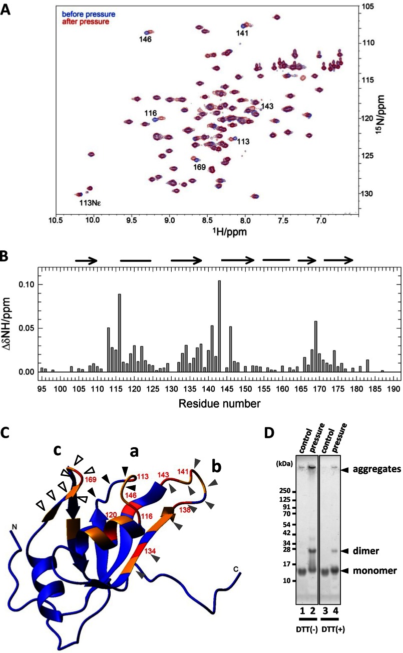
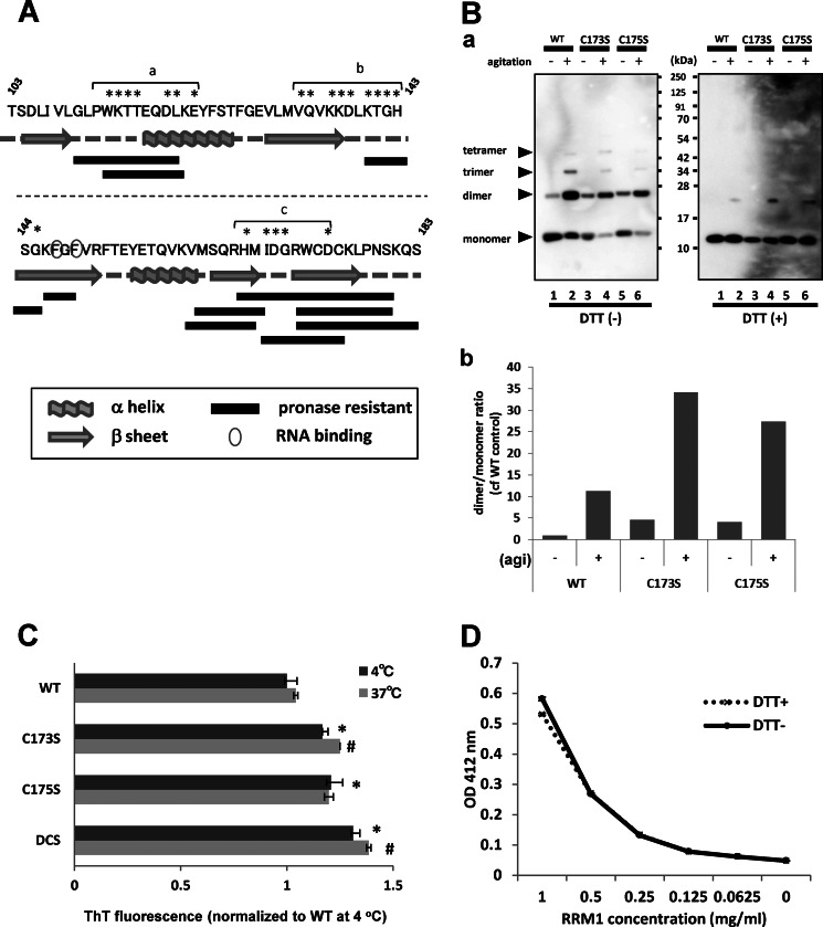
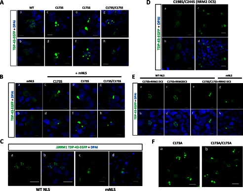
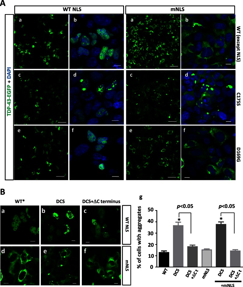
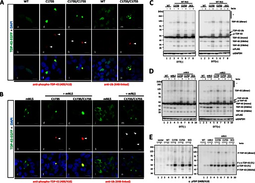
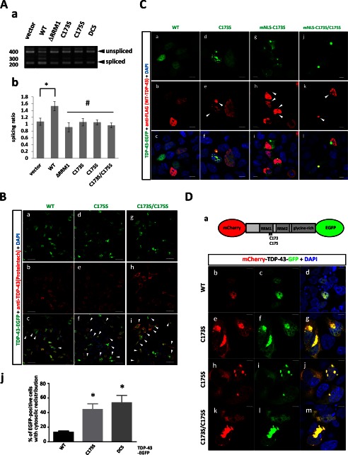
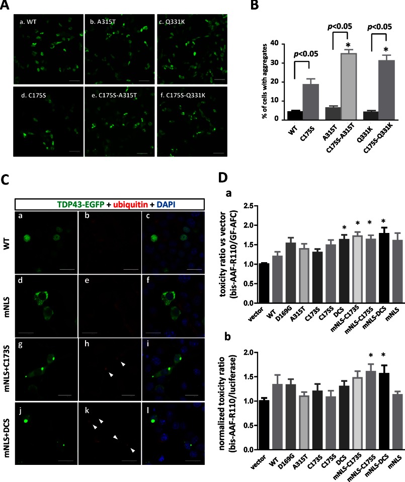
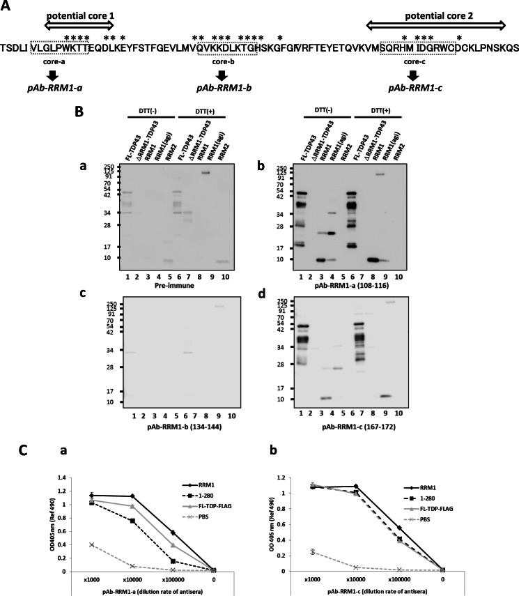
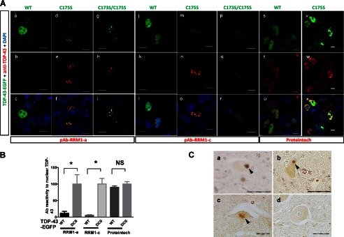
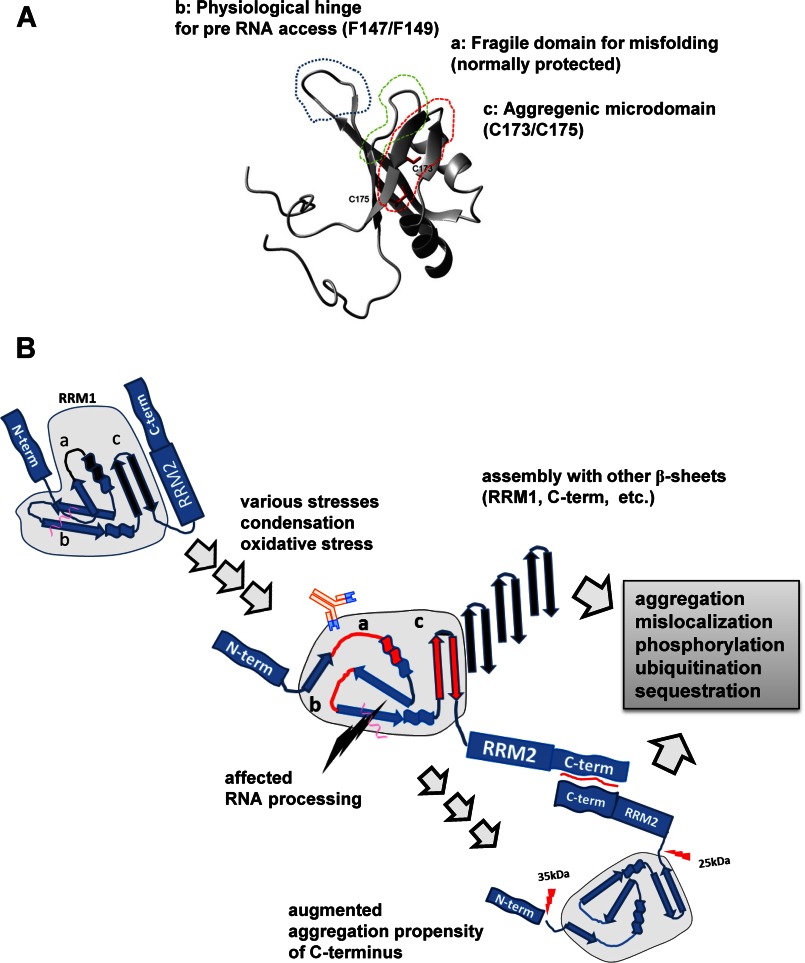
Aggregation of TAR DNA-binding protein of 43 kDa (TDP-43) is a pathological signature of amyotrophic lateral sclerosis (ALS). Although accumulating evidence suggests the involvement of RNA recognition motifs (RRMs) in TDP-43 proteinopathy, it remains unclear how native TDP-43 is converted to pathogenic forms. To elucidate the role of homeostasis of RRM1 structure in ALS pathogenesis, conformations of RRM1 under high pressure were monitored by NMR. We first found that RRM1 was prone to aggregation and had three regions showing stable chemical shifts during misfolding. Moreover, mass spectrometric analysis of aggregated RRM1 revealed that one of the regions was located on protease-resistant β-strands containing two cysteines (Cys-173 and Cys-175), indicating that this region served as a core assembly interface in RRM1 aggregation. Although a fraction of RRM1 aggregates comprised disulfide-bonded oligomers, the substitution of cysteine(s) to serine(s) (C/S) resulted in unexpected acceleration of amyloid fibrils of RRM1 and disulfide-independent aggregate formation of full-length TDP-43. Notably, TDP-43 aggregates with RRM1-C/S required the C terminus, and replicated cytopathologies of ALS, including mislocalization, impaired RNA splicing, ubiquitination, phosphorylation, and motor neuron toxicity. Furthermore, RRM1-C/S accentuated inclusions of familial ALS-linked TDP-43 mutants in the C terminus. The relevance of RRM1-C/S-induced TDP-43 aggregates in ALS pathogenesis was verified by immunolabeling of inclusions of ALS patients and cultured cells overexpressing the RRM1-C/S TDP-43 with antibody targeting misfolding-relevant regions. Our results indicate that cysteines in RRM1 crucially govern the conformation of TDP-43, and aberrant self-assembly of RRM1 at amyloidogenic regions contributes to pathogenic conversion of TDP-43 in ALS.

Keywords: Amyotrophic Lateral Sclerosis (Lou Gehrig's Disease); Cell Biology; Protein Chemical Modification; Protein Misfolding; Structural Biology; TAR DNA-binding Protein 43 kDa.


Figures

FIGURE 1.
FIGURE 1.
Pressure-induced structural changes in RRM1. A, overlaid 15N/1H HSQC spectra for the stable isotope-labeled RRM1 domain at 30 bars and 25 °C before (blue) and after (red) pressure treatment. Residues showing large chemical shift changes are indicated by the residue number. B, deviations of 1H chemical shifts in RRM1. ΔδNH was calculated by the equation, ((ΔδH)2 + (ΔδN/5)2)0.5. The α-helix and β-sheet are indicated at the top by lines and arrows, respectively. C, three-dimensional structure of RRM1 in solution as determined by NMR. Residues showing large chemical shifts are indicated by arrowheads; colors indicate the degree of shift (0.02 < Δδ < 0.03 ppm (orange) and 0.03 < Δδ ppm (red)). Three clusters of amino acid residues showing irreversible chemical shifts on NMR are indicated by arrowheads: core-a (a, black), core-b (b, gray), and core-c (c, white). D, SDS-polyacrylamide gel showing the SDS-resistant dimer of the a-, b-, and c-domains following exposure to 2000-bar pressure. Bands (kDa) corresponding to the monomer and dimer proteins are indicated by arrows.
FIGURE 2.
FIGURE 2.
Native RRM1 is a soluble monomer that is prone to aggregation. A, seeding effect of RRM1 aggregates assessed with disulfide-mediated dimers. The RRM1 proteins were agitated for 16 h or non-treated (ctrl), followed by postincubation at 4 °C for 1, 5, 7, or 14 days, and total protein mixture was separated by non-reducing SDS-PAGE. a, Coomassie Brilliant Blue staining; b, a densitometry for the ratio of the dimer to the monomer. B, gel filtration analysis showing the absorbance at 215 nm of RRM1 with (closed circles) or without (open circles) agitation. Agitation markedly reduces the amount of monomeric RRM1 in solution with no emergence of oligomers. Molecular size markers are ovalbumin (43 kDa), copper zinc superoxide dismutase (32 kDa), myoglobin (17.6 kDa), and aprotinin (6.5 kDa). C, Coomassie Brilliant Blue staining of a PFO-polyacrylamide gel (see "Experimental Procedures") for the RRM1 domain after transient agitation or the application of heat stress. Single, double, and triple arrowheads indicate the monomer, dimer, and trimer sizes of RRM1, respectively. Asterisks indicate the high molecular weight species of RRM1. D, thioflavin T (ThT) fluorescence assay showing time-dependent amyloid formation of agitated RRM1. Recombinant WT RRM1 was incubated with BTA-1 at either 24 h or 7 days after 16-h agitation or static incubation at room temperature (RT) as a control. *, p < 0.05 versus control sample by one-way ANOVA of Bonferroni's test. Each value is expressed as ThT ratio to room temperature treatment with a 24-h postincubation (n = 3; ±S.E. (error bars)). E, atomic force microscope analysis of RRM1 aggregation after a 16-h agitation. Images show proteins on days 0 (a), 5 (b), and 7 (c and d). Large globular particles and fibrillar aggregates, indicated by arrowheads were present after agitation. F, a, schematic illustration of human TDP-43 with domain information. b, non-reducing SDS-PAGE analysis of RRM1, RRM2, and N-terminal fragments spanning RRM1 (aa 1–183) and RRM2 (aa 1–265), with or without overnight agitation. Protein solutions were separated into supernatant and pellet fractions after a 7-day incubation at 4 °C after the agitation. *, dimer forms of each protein (RRM1, aa 1–183 fragment, and aa 1–265 fragment).
FIGURE 3.
FIGURE 3.
The interaction of RRM1 with TG-repeated oligonucleotides did not influence the aggregate formation of RRM1. A, size exclusion chromatography showing the interaction between RRM1 and oligonucleotides comprising 12 repeats of thymine-guanine, (TG)12. The figure shows that peak flows for the mixture of RRM1 and (TG)12 merged into a single fraction, corresponding to the combined molecular weight of both, whereas individual applications led to different and smaller fractions. B, SDS-polyacrylamide gels showing recombinant RRM1 proteins (40 μm) incubated with (TG)12, adenine-cytosine (AC)12 (40 μm), or PBS and subjected to 16-h agitation at room temperature. Samples with (+) or without (−) DTT were separated by SDS-PAGE and stained using Coomassie Brilliant Blue. No alteration of dimer formation was observed in the presence of (TG)12. Single and double asterisks indicate RRM1 monomer and dimer, respectively.
FIGURE 4.
FIGURE 4.
Amyloid formation by RRM1 via specific β-sheet assembly by cysteine substitutions. A, schematic illustration of RRM1 residues; protease-resistant fragments and secondary structures are indicated by solid bars and gray schematics, respectively. Pronase-resistant fragments of aggregated RRM1 were analyzed by LC-MS/MS; regions with monoisotopic m/z > 800 are shown. The amino acid residues indicated by asterisks showed irreversible changes in NMR chemical shift after 2000-bar pressure treatment, which were designated as misfolding-relevant core-a, -b, and -c. B, a, Western blot analysis of WT or single RRM1-C/S mutants (C173S or C175S) under reducing (DTT (+)) or non-reducing (DTT (−)) conditions. Proteins were incubated for 7 days at 4 °C after 16-h agitation (+) or non-treatment (−). b, densitometric analysis of the dimer/monomer ratio, indicating that the single C/S mutation accelerates aggregate formation containing an intermolecular disulfide bond. agi, agitation. C, ThT fluorescence assay showing that RRM1 aggregates form amyloid fibrils. Recombinant RRM1 proteins (0.03 mm) were reacted with a ThT derivative (BTA-1) after 24-h incubation at 4 or 37 °C. Data represent the -fold increase in the ThT ratio compared with the controls. Each value expresses mean ± S.E. (n = 3). *, p < 0.05 versus control sample by one way ANOVA of Bonferroni's test. D, free state of cysteine residues in the de novo RRM1 protein. Absorbance (412 nm) values obtained from the Ellman's test using 5,5′-dithiobis(2-nitrobenzoic acid). RRM1 proteins at the indicated concentrations were incubated with 5,5′-dithiobis(2-nitrobenzoic) acid for 20 min at 22 °C. Data are expressed as the average of duplicate values. RRM1 proteins either with (+) or without (−) DTT effectively reacted with 5,5′-dithiobis(2-nitrobenzoic) acid, indicating the free thiol moieties.
FIGURE 5.
FIGURE 5.
RRM1-C/S substitutions induced cytoplasmic and nuclear inclusions of TDP-43. Shown are confocal analyses of HEK293A cells transiently transfected with EGFP-fused TDP-43. A and B, RRM1-C/S mutations in TDP-43 induced aggregates in the nucleus and cytosol. Cells were transiently transfected with full-length WT or RRM1-C/S of TDP-43-EGFP (green) with WT (A) or modified (B) NLS. Scale bar, 10 μm. C, the RRM1 deletion mutant of TDP-43 formed marked nuclear inclusions, but RRM1-deleted TDP-43 with modified NLS did not. HEK293A cells were transiently transfected with EGFP-fused TDP-43 lacking RRM1 with WT (a and b) or modified (c and d) NLS. Scale bar, 10 μm. D, RRM2-C/S had no impact on the TDP-43 aggregation. Cells were transfected with full-length WT or RRM2-C/S double mutant (RRM2 DCS) TDP-43-EGFP (green). Scale bars, 10 μm (a and b) and 30 μm (c and d). E, disulfide bonding in RRM1 and RRM2 is not inevitable for TDP-43 aggregation. Cells were transfected with RRM1 single mutants and the RRM2-C/S double mutant (RRM2 DCS) of TDP-43-EGFP (green). Double C/S substitutions at Cys-198/Cys-244 (RRM2 DCS) did not affect nuclear or cytosolic TDP-43 inclusions caused by C/S mutations at RRM1. Scale bar, 10 μm. F, C/A mutants in Cys-173 and/or C175S showed the same aggregation as C/S mutants. Scale bar, 10 μm. Nuclei were stained by DAPI (blue).
FIGURE 6.
FIGURE 6.
Crucial roles of cysteines in RRM1 and C-terminal domain in the formation of cytosolic inclusions of TDP-43. A, sporadic ALS mutation D169G mutation in RRM1 domain caused no TDP-43 aggregation. Confocal analysis of HEK293A cells overexpressing nuclear (A) or mislocalized (mNLS) (B) TDP-43-EGFP (green), with C175S (c and d) or D169G (e and f) mutations. Scale bar, 10 mm (a, c, and e) or 50 μm (b, d, and f). B, C terminus mediates RRM1-C/S-induced aggregation of TDP-43. Confocal analysis of HEK293A cells transiently transfected with nuclear (a–c, WT NLS) or cytosolic (d–f, mNLS) TDP-43-EGFP carrying the double C173S/C175S mutation (DCS), either with (c and f) or without (b and e) the C terminus (aa 266–414, ΔCt). WT*, WT NLS. g, quantification of the effect of the C-terminal tail of TDP-43 on nuclear or cytosolic aggregates caused by RRM1-C/S mutation. The percentage of aggregate holding cells in the total transfected cells was obtained by counting. *, p < 0.05 versus nuclear or cytosolic TDP-43 without any C/S mutation (WT or mNLS). †, p < 0.05 between DCS mutants with and without C-terminal deletions by one-way ANOVA of Bonferroni's test. Data represent the mean ± S.E. (error bars) (n = 6).
FIGURE 7.
FIGURE 7.
TDP-43 inclusions with RRM1-C/S mutation were phosphorylated, ubiquitinated, and disulfide-free. A and B, confocal analysis of SHSY-5Y cells transfected with full-length WT or RRM1-C/S mutants of TDP-43-EGFP (green). Cells were immunostained with antibodies (red) targeting phospho-TDP-43 at Ser-409/Ser-410 (a–i) or Lys-48-linked ubiquitin (j–o). In B, all constructs contained mNLS. Nuclei were stained by DAPI (blue). Phosphorylated or ubiquitinated inclusions in the cytoplasm are indicated by arrowheads. Note that cytoplasmic inclusions (green) are strongly immunoreactive to both antibodies compared with nuclear aggregates. Scale bar, 10 μm. C and D, Western blots showing that disulfide-irrelevant oligomers of TDP-43 harboring RRM1-C/S substitutions under the proteasome inhibitor lactacystin are more prominent in the cytosol (D) than in the nucleus (C). TDP-43-FLAG was overexpressed in HEK293A cells. Cell lysates were analyzed by SDS-PAGE and incubated with antibodies targeting FLAG and GAPDH. Note that DTT marginally reduced oligomerization (indicated by an asterisk) and increased dimer formation in C173S and C175S mutants. E, Western blot of TDP-43 with RRM1-C/S substitutions using an antibody targeting TDP-43 phosphorylated at Ser-409/Ser-410. TDP-43 with mNLS is phosphorylated to a greater extent than WT, which is enhanced by lactacystin (right, lanes 3 and 4). Conversely, TDP-43 harboring the RRM1-C/S substitution is more strongly phosphorylated, and modification of NLS or lactacystin treatment augmented the phosphorylated TDP-43 (both left and right, lanes 5–10).
FIGURE 8.
FIGURE 8.
C/S substitution in RRM1 induces diverse dysfunctions of ALS-linked TDP-43 proteinopathies. A, the exon 9 skipping assay for cystic fibrosis transmembrane conductance regulator RNA. cDNA from HEK293A cells was analyzed 48 h after co-transfection with TDP-43-EGFP and TG13T5 minigene reporter plasmids. a and b, which show the agarose gel image and densitometric analysis, respectively, show the impaired RNA splicing effect of the WT and mutant TDP-43-EGFP proteins, and the ratio of sliced to unspliced fragments was obtained. Data represent the mean ± S.E. n = 4. *, p < 0.05 versus vector control; #, p < 0.05 versus WT TDP-43 by one-way ANOVA of Bonferroni's test. B, TDP-43 with RRM1-C/S substitutions induces cytosolic mislocalization of TDP-43. HEK293A cells were transiently transfected with WT (a–c) or RRM1-C/S mutant TDP-43-EGFP (d–i) and were analyzed using rabbit polyclonal anti-TDP-43 antibody. Scale bar, 50 μm. j, quantification of TDP-43-EGFP-expressing cells with cytosolic mislocalized TDP-43. Data represent the mean ± S.E. (n = 4). In each experiment, 21–70 cells were counted. *, p < 0.05 versus WT by one-way ANOVA of Bonferroni's test. C, immunofluorescence analysis showing that nuclear or cytosolic aggregates of TDP-43 with RRM1-C/S substitution recruit nuclear WT TDP-43. HEK293A cells were co-transfected with the nuclear (a–f) or cytosolic (g–l) forms of WT or C/S mutant TDP-43-EGFP (green) together with FLAG-tagged WT TDP-43. Cells were stained with an anti-FLAG antibody (red). Colocalized TDP-43-EGFP and TDP-43-FLAG are indicated by arrowheads. Scale bar, 10 μm. D, full-length TDP-43 is redistributed into the cytosol by RRM1-C/S substitution. a, schematic illustration of mCherry-TDP-43-GFP. 5′- and 3′- wild-type and mutant TDP-43 genes were fused with mCherry and GFP, respectively, in an in-frame manner. b–m, co-localization of both N and C termini of TDP-43 containing C173S and/or C175S substitutions. HEK-293A cells were transfected with the indicated constructs and were cultured for 32 h. The transfected cells were further treated with 10 mm lactacystin for 16 h, followed by observation with a confocal microscope. Scale bar, 10 μm.
FIGURE 9.
FIGURE 9.
RRM1-C/S mutations unravel the pathogenic properties of TDP-43. A, RRM1-C/S substitution enhances the aggregation propensity of TDP-43 with familial ALS-linked mutations in the C-terminal domain. EGFP-fused WT or A315T and Q331K mutants of TDP-43, either with (d–f) or without (a–c) the C175S mutation, were transiently expressed in HEK293A cells. B, quantification of cells with aggregates. Data represent the mean percentages of cells harboring aggregates in total transfected cells ± S.E. (n = 8 images); 20–120 cells were counted in each image. *, p < 0.05 by one-way ANOVA of Bonferroni's test. C, cytosolic aggregates of TDP-43 with RRM1-C/S injure the motor neuron cell line. a–l, confocal micrographs showing subcellular distributions and inclusion formation of TDP-43-EGFP in NSC34. D, a and b, fluorometric assay for neurotoxicity of RRM1-C/S mutants in NSC-34 cells. Murine motor neuron NSC34 cells plated in a 96-well culture plate were transiently transfected with plasmids expressing TDP-43-FLAG (0.2 μg/well) of WT, ALS-linked mutants (D169G and A315T), or Cys-173/Cys-175 mutants (C173S, C175S, and C173S/C175S (DCS)) with or without mNLS. Data represent the mean ± S.E. (n = 12 wells from 4 independent experiments; each value is normalized to vector control in each experiment). *, p < 0.05 versus vector control by one-way ANOVA of Bonferroni's test. b, dead cell marker bis-AAF-R110 was also normalized to luciferase activity from co-transfected Renilla luciferase reporter plasmid. The Renilla luciferase reporter gene (0.02 μg/well) was co-transfected with pcDNA-TDP-43-FLAG. Data represent the mean ± S.E. (n = 11–12). Each value is normalized to vector control in each experiment). *, p < 0.05 versus vector control by one-way ANOVA of Dunnett's test.
FIGURE 10.
FIGURE 10.
Evaluation of polyclonal antibody for the detection of aggregation-relevant cores. A, schematic illustration showing the antigenic sequences in RRM1 used to generate the polyclonal antibodies raised against core-a (aa 108–116), core-b (aa 134–142), and core-c (aa 163–173), which are designated as pAb RRM1-a, -b, and -c, respectively. Asterisks indicate the residues that showed irreversible chemical shifts on NMR. B, Western blot analysis using antibody against three aggregation-relevant domains (Ab1, Ab2, and Ab3) in the presence (DTT (+)) and absence (DTT (−)) of DTT. The antibody 1 (Ab1; against aa 108–116) and the antibody 3 (Ab3; against aa 167–172) recognize RRM1 and full-length recombinant TDP-43 but not RRM2 or the RRM1 deletion mutant of TDP-43. C, ELISA study showing the immunoreactivity of pAb RRM1-a (a) and pAb RRM1-c (b) against recombinant human TDP-43 proteins of various types.
FIGURE 11.
FIGURE 11.
Misfolding-relevant core in RRM1 is an immunogenic marker of TDP-43 inclusions. A, confocal micrographs showing SHSY-5Y cells transiently expressing EGFP-fused WT or C/S mutant TDP-43. Cells were immunostained with the antibody targeting core-a (a–i; pAb RRM1-a) or core-c (j–r; pAb RRM1-c) or a commercially available anti-TDP-43 antibody (s–x; Proteintech). The bottom panels show overlaid images of the EGFP (green) and antibody (red) signals. Nuclei were stained with DAPI (blue). Scale bar, 10 μm. B, quantification of the aggregate-specific reactivity of the pAb RRM1-a and pAb RRM1-c antibodies, compared with that of a commercially available anti-TDP-43 antibody (Proteintech). The fluorescence of EGFP and the antibody was measured in each nucleus expressing WT or C173S/C175S mutant (DCS) TDP-43. The ratio of antibody to EGFP fluorescence was determined; WT data are normalized to the DCS data, which are expressed as Ab reactivity to nuclear TDP-43. Data represent the mean ± S.E. (n = 15–117 nuclei). *, p < 0.05 by one-way ANOVA of Bonferroni's test. C, representative immunohistochemical photographs of spinal cord sections from three sporadic ALS patients (a–c) and a subject with myasthenia gravis (d) were stained with antibodies targeting RRM1-a. Arrowheads indicate Lewy body-like hyaline inclusions (a and b) and a skein-like inclusion (c). Scale bar, 20 μm.
FIGURE 12.
FIGURE 12.
Hypothetical roles of misfolding-relevant cores in RRM1. A, ribbon structures showing the locations of misfolding-relevant RRM1 core-a, -b, and -c. B, hypothetical scheme of TDP-43 proteinopathy development. For clarity, RRM1 is indicated by a shaded area; core-a, -b, and -c are also indicated. These cores may not be exposed under physiological conditions. Various cellular stresses, such as oxidative stress or regional condensation of TDP-43, may alter the β-sheet assembly, which may induce transient disulfide bonds, ultimately resulting in pathological TDP-43 proteinopathies with the participation of the C terminus.

References

    1. Arai T., Hasegawa M., Akiyama H., Ikeda K., Nonaka T., Mori H., Mann D., Tsuchiya K., Yoshida M., Hashizume Y., Oda T. (2006) TDP-43 is a component of ubiquitin-positive tau-negative inclusions in frontotemporal lobar degeneration and amyotrophic lateral sclerosis. Biochem. Biophys. Res. Commun. 351, 602–611 - PubMed
    1. Neumann M., Sampathu D. M., Kwong L. K., Truax A. C., Micsenyi M. C., Chou T. T., Bruce J., Schuck T., Grossman M., Clark C. M., McCluskey L. F., Miller B. L., Masliah E., Mackenzie I. R., Feldman H., Feiden W., Kretzschmar H. A., Trojanowski J. Q., Lee V. M. (2006) Ubiquitinated TDP-43 in frontotemporal lobar degeneration and amyotrophic lateral sclerosis. Science 314, 130–133 - PubMed
    1. Baloh R. H. (2012) How do the RNA-binding proteins TDP-43 and FUS relate to amyotrophic lateral sclerosis and frontotemporal degeneration, and to each other? Curr. Opin. Neurol. 25, 701–707 - PubMed
    1. Pesiridis G. S., Lee V. M., Trojanowski J. Q. (2009) Mutations in TDP-43 link glycine-rich domain functions to amyotrophic lateral sclerosis. Hum. Mol. Genet. 18, R156–R162 - PMC - PubMed
    1. Gendron T. F., Josephs K. A., Petrucelli L. (2010) Review. Transactive response DNA-binding protein 43 (TDP-43). Mechanisms of neurodegeneration. Neuropathol. Appl. Neurobiol. 36, 97–112 - PMC - PubMed

Publication types

MeSH terms

Cite

AltStyle によって変換されたページ (->オリジナル) /