This site needs JavaScript to work properly. Please enable it to take advantage of the complete set of features!
Skip to main page content
U.S. flag

An official website of the United States government

Dot gov

The .gov means it’s official.
Federal government websites often end in .gov or .mil. Before sharing sensitive information, make sure you’re on a federal government site.

Https

The site is secure.
The https:// ensures that you are connecting to the official website and that any information you provide is encrypted and transmitted securely.

NIH NLM Logo
Log in
Access keys NCBI Homepage MyNCBI Homepage Main Content Main Navigation
. 2013 Jul;18(4):495-501.
doi: 10.1007/s12192-013-0405-3. Epub 2013 Feb 3.

ERK signaling is triggered by hepatitis C virus E2 protein through DC-SIGN

Affiliations

ERK signaling is triggered by hepatitis C virus E2 protein through DC-SIGN

Lan-Juan Zhao et al. Cell Stress Chaperones. 2013 Jul.

Abstract

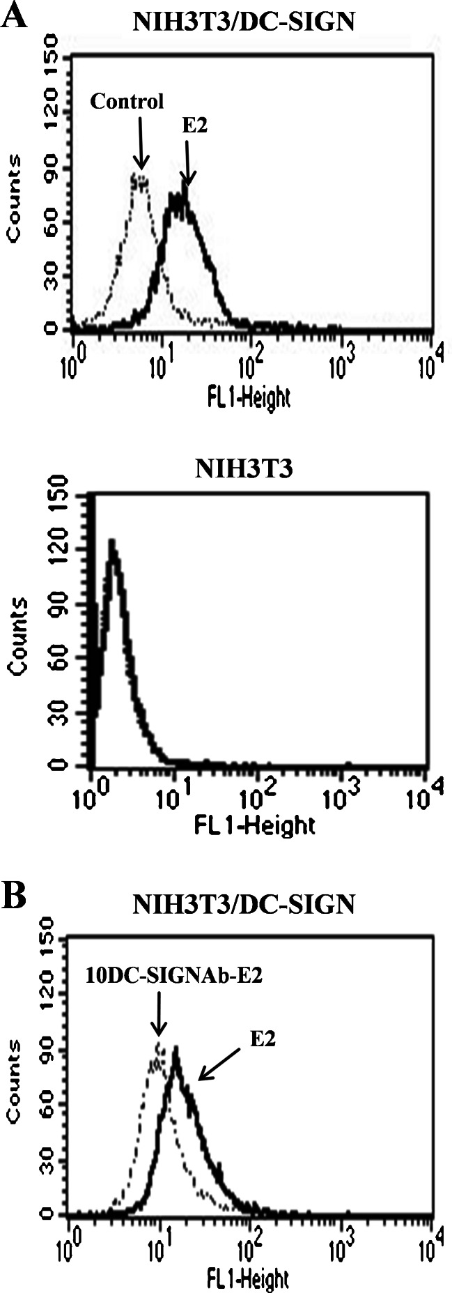
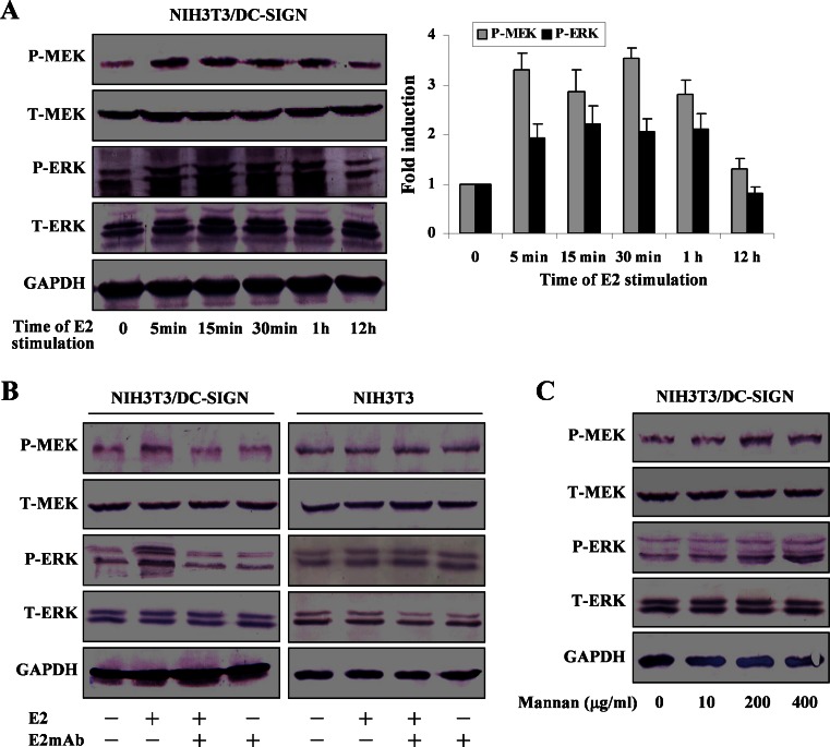
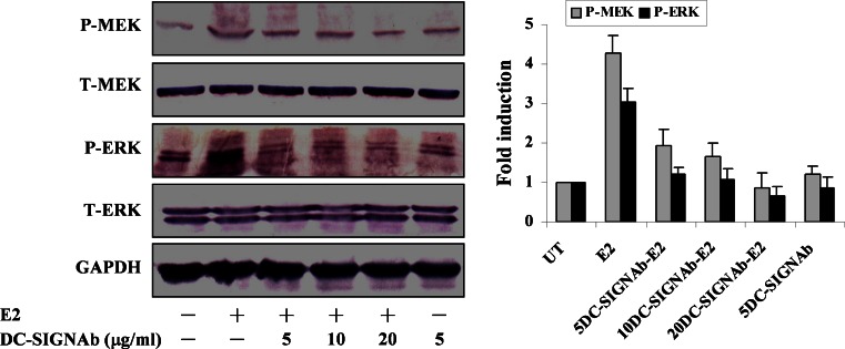
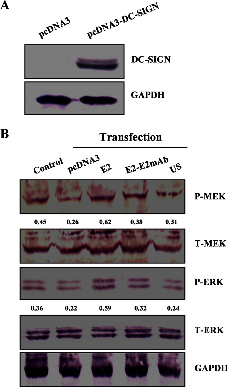
Dendritic cell-specific intercellular adhesion molecule-3-grabbing nonintegrin (DC-SIGN) is a binding receptor for hepatitis C virus (HCV). Binding of HCV envelope protein E2 to target cells is a prerequisite to DC-SIGN-mediated signaling. Using cell lines with stable or transient expression of DC-SIGN, we investigated effects of soluble HCV E2 protein on ERK pathway. MEK and ERK are activated by the E2 in NIH3T3 cells stably expressing DC-SIGN. Treatment of the cells with antibody to DC-SIGN results in inhibition of the E2 binding as well as the E2-induced MEK and ERK activation. In HEK293T cells transiently expressing DC-SIGN, activation of MEK and ERK is also induced by the E2. Activation of ERK pathway by HCV E2 through DC-SIGN provides useful information for understanding cellular receptor-mediated signaling.


Figures

Fig. 1
Fig. 1
HCV E2 binding to NIH3T3/DC-SIGN via DC-SIGN. a NIH3T3/DC-SIGN and NIH3T3 cells were incubated with the E2 and the E2 binding was detected by flow cytometry using goat anti-E2 Ab and Fluorescein isothiocyanate-conjugated rabbit anti-goat IgG. Cells were stained with the goat anti-E2 Ab and the secondary antibody as control. b NIH3T3/DC-SIGN cells were pre-incubated with anti-DC-SIGN mAb prior to the E2 incubation. The E2 binding was analyzed as above. Similar results were obtained in three independent experiments, and one representative experiment is shown
Fig. 2
Fig. 2
Activation of ERK pathway by HCV E2. a NIH3T3/DC-SIGN cells were cultured with the E2 for the indicated time periods. Cell lysates were prepared for assessment of total (T-) and phosphorylated (P-) MEK and ERK by Western blotting using antibodies against total or phosphorylated kinases. Quantification was made using GeneTools software, and results correspond to the ratio between the amount of phosphorylated MEK or ERK and the amount of total MEK or ERK normalized to the control without E2 stimulation. Results represent mean and SD of three experiments. b NIH3T3/DC-SIGN and NIH3T3 cells were treated with the E2 or the E2-E2 mAb. c NIH3T3/DC-SIGN cells were treated with mannan at the indicated concentrations. Total and phosphorylated MEK and ERK were analyzed by Western blotting. Representative results out of four experiments are shown
Fig. 3
Fig. 3
Effect of DC-SIGN mAb on MEK and ERK phosphorylation induced by HCV E2. NIH3T3/DC-SIGN cells were treated with anti-DC-SIGN mAb at the indicated concentrations before treatment with the E2. Total (T-) and phosphorylated (P-) MEK and ERK were analyzed by Western blotting. Quantification was made using GeneTools software, and results correspond to the ratio between the amount of phosphorylated MEK or ERK and the amount of total MEK or ERK normalized to the untreated control (UT). Results represent mean and SD of three experiments
Fig. 4
Fig. 4
Phosphorylation of MEK and ERK in HEK293T transiently expressing DC-SIGN under HCV E2 stimulation. a HEK293T cells were transfected with pcDNA3-DC-SIGN expression plasmid or pcDNA3 vector, and DC-SIGN expression was detected by Western blotting. b The transfected cells were stimulated with the E2 or the E2-E2 mAb. Total (T-) and phosphorylated (P-) kinases were analyzed by Western blotting. Changes in the amount of phosphorylated MEK or ERK over the amount of total MEK or ERK are shown below each blot. Control untransfected and unstimulated, US pcDNA3-DC-SIGN transfection without the E2 stimulation. Representative results out of three experiments are shown

References

    1. Alvarez CP, Lasala F, Carrillo J, Muñiz O, Corbí AL, Delgado R. C-type lectins DC-SIGN and L-SIGN mediate cellular entry by Ebola virus in cis and in trans. J Virol. 2002;76:6841–6844. doi: 10.1128/JVI.76.13.6841-6844.2002. - DOI - PMC - PubMed
    1. Balasubramanian A, Ganju RK, Groopman JE. Hepatitis C virus and HIV envelope proteins collaboratively mediate interleukin-8 secretion through activation of p38 MAP kinase and SHP2 in hepatocytes. J Biol Chem. 2003;278:35755–35766. doi: 10.1074/jbc.M302889200. - DOI - PubMed
    1. Balasubramanian A, Koziel M, Groopman JE, Ganju RK. Molecular mechanism of hepatic injury in coinfection with hepatitis C virus and HIV. Clin Infect Dis. 2005;41(Suppl 1):S32–37. doi: 10.1086/429493. - DOI - PubMed
    1. Brazzoli M, Bianchi A, Filippini S, Weiner A, Zhu Q, Pizza M, Crotta S. CD81 is a central regulator of cellular events required for hepatitis C virus infection of human hepatocytes. J Virol. 2008;82:8316–8329. doi: 10.1128/JVI.00665-08. - DOI - PMC - PubMed
    1. Caparrós E, Munoz P, Sierra-Filardi E, Serrano-Gómez D, Puig-Kröger A, Rodríguez-Fernández JL, Mellado M, Sancho J, Zubiaur M, Corbí AL. DC-SIGN ligation on dendritic cells results in ERK and PI3K activation and modulates cytokine production. Blood. 2006;107:3950–3958. doi: 10.1182/blood-2005年03月12日52. - DOI - PubMed

Publication types

MeSH terms

Substances

LinkOut - more resources

Cite

AltStyle によって変換されたページ (->オリジナル) /