This site needs JavaScript to work properly. Please enable it to take advantage of the complete set of features!
Skip to main page content
U.S. flag

An official website of the United States government

Dot gov

The .gov means it’s official.
Federal government websites often end in .gov or .mil. Before sharing sensitive information, make sure you’re on a federal government site.

Https

The site is secure.
The https:// ensures that you are connecting to the official website and that any information you provide is encrypted and transmitted securely.

NIH NLM Logo
Log in
Access keys NCBI Homepage MyNCBI Homepage Main Content Main Navigation
. 2012 Dec 1;189(11):5336-46.
doi: 10.4049/jimmunol.1201028. Epub 2012 Oct 24.

Prolonged neutrophil dysfunction after Plasmodium falciparum malaria is related to hemolysis and heme oxygenase-1 induction

Affiliations

Prolonged neutrophil dysfunction after Plasmodium falciparum malaria is related to hemolysis and heme oxygenase-1 induction

Aubrey J Cunnington et al. J Immunol. .

Abstract

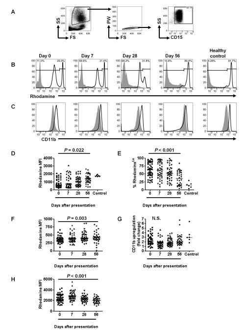
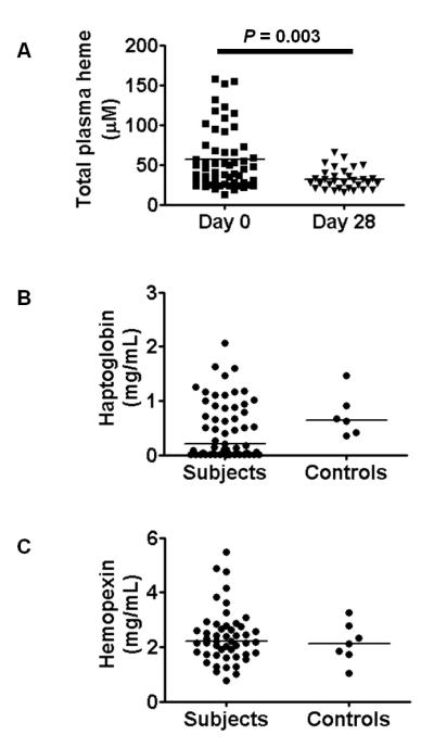
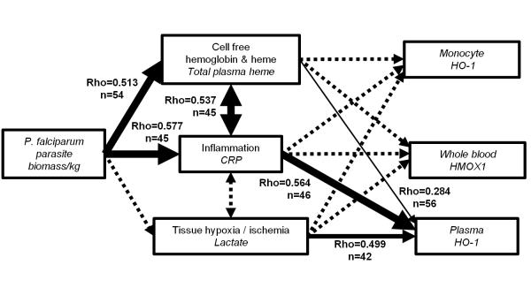
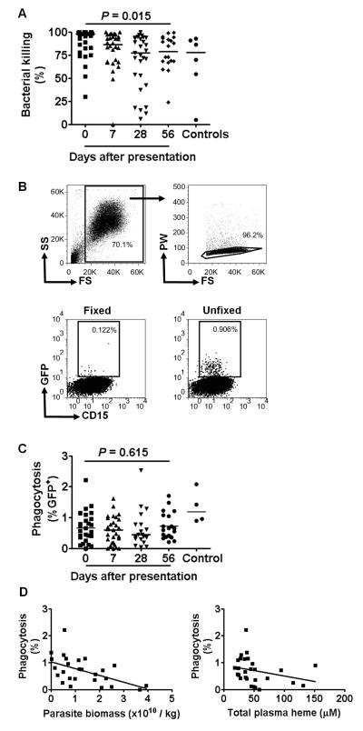
It is not known why people are more susceptible to bacterial infections such as nontyphoid Salmonella during and after a malaria infection, but in mice, malarial hemolysis impairs resistance to nontyphoid Salmonella by impairing the neutrophil oxidative burst. This acquired neutrophil dysfunction is a consequence of induction of the cytoprotective, heme-degrading enzyme heme oxygenase-1 (HO-1) in neutrophil progenitors in bone marrow. In this study, we assessed whether neutrophil dysfunction occurs in humans with malaria and how this relates to hemolysis. We evaluated neutrophil function in 58 Gambian children with Plasmodium falciparum malaria [55 (95%) with uncomplicated disease] and examined associations with erythrocyte count, haptoglobin, hemopexin, plasma heme, expression of receptors for heme uptake, and HO-1 induction. Malaria caused the appearance of a dominant population of neutrophils with reduced oxidative burst activity, which gradually normalized over 8 wk of follow-up. The degree of neutrophil impairment correlated significantly with markers of hemolysis and HO-1 induction. HO-1 expression was increased in blood during acute malaria, but at a cellular level HO-1 expression was modulated by changes in surface expression of the haptoglobin receptor (CD163). These findings demonstrate that neutrophil dysfunction occurs in P. falciparum malaria and support the relevance of the mechanistic studies in mice. Furthermore, they suggest the presence of a regulatory pathway to limit HO-1 induction by hemolysis in the context of infection and indicate new targets for therapeutic intervention to abrogate the susceptibility to bacterial infection in the context of hemolysis in humans.


Figures

FIGURE 1
FIGURE 1
P. falciparum malaria causes prolonged impairment of the neutrophil oxidative burst. (A) Representative FACS plots showing the gating of the neutrophil population based on forward scatter and side scatter characteristics followed by selection of single cells based on pulse width and forward scatter, and selection of the CD15+ population. (B,C) Rhodamine (B) and CD11b (C) fluorescence of unstimulated (filled histogram) and PMA-stimulated neutrophils (unfilled histogram) on days 0, 7, 28 and 56 after presentation with P. falciparum malaria. Representative plots from a healthy control child are also shown for comparison. RhodamineHi and rhodamineLo populations of neutrophils were defined for each sample by partition at the nadir of the bimodal distribution, and percentages of the total cells in each population are shown (B). (D-H) Longitudinal analyses of neutrophil function, compared using Friedman’s two way ANOVA for all subjects with valid data at all time points. Healthy controls are also shown for comparison, but not included in the statistical analysis (D,E,G). Horizontal lines represent medians. n stated for valid data at every time point. (D) Rhodamine MFI for all neutrophils (rhodamineHi and rhodamineLo considered together as a single population); n=32. (E) Proportion of neutrophils that are rhodamineLo; n=28. (F) Rhodamine MFI of rhodamineLo neutrophils; n=28. (G) Degranulation of all neutrophils, assessed by fold change in surface CD11b (PMA stimulated CD11b MFI: unstimulated CD11b MFI); n=29. (H) Rhodamine MFI of rhodamineHi neutrophils; n=32.
FIGURE 2
FIGURE 2
Indicators of hemolysis in subjects with P. falciparum malaria. (A) Total plasma heme on day 0 and day 28, compared using Wilcoxon matched pairs test for those with data at both time points (n=32). (B) Distribution of plasma haptogobin levels at day 0, n=57. For comparison levels in six healthy control children are also shown. (C) Distribution of plasma hemopexin levels at day 0, n=49. For comparison levels in six healthy control children are also shown.
FIGURE 3
FIGURE 3
Factors associated with heme oxygenase-1 expression in P. falciparum malaria. (A-C): heme oxygenase-1 induction. (A) Representative FACS analysis of HO-1 induction in moncoytes showing exclusion of red blood cells, gating on single cells, and subsequent definition of monocytes as CD14+. Histograms show fluorescence of monocytes stained intracellularly with control antibody (filled) or anti-HO-1 antibody (unfilled) followed by a secondary detection antibody. Quantitative data for HO-1 expression in monocytes (ratio of anti-HO-1 to control antibody fluorescence) for all subjects on days 0, 7 and 28 are shown in the right hand panel, compared using Friedman’s two way ANOVA (n=20). (B) The distribution of Plasma HO-1 on day 0, n=57. For comparison levels in six healthy control children are also shown. (C) Whole blood HMOX1 RNA expression, measured by qRT-PCR, compared between samples on day 0 and day 28 using Wilcoxon matched pairs test for those with data at both time points (n=42). (D-H) CD163 and CD91 expression on monocytes and neutrophils. (D) Representative flow cytometry analysis of healthy control subject neutrophils (CD14CD16b+) and monocytes (CD14+CD16b), showing staining with respective control (filled histogram) and anti-CD91 or anti-CD163 antibodies (unfilled histograms). Quantitative data for expression (ratio to control antibody fluorescence) of CD163 in monocytes (E) and neutrophils (F), and CD91 in monocytes (G) and neutrophils (H), compared using Friedman’s two way ANOVA for all subjects with valid data at all time points (CD163, n=19; CD91 n=20). Horizontal bars represent medians. n stated for those with valid data at all time points.
FIGURE 4
FIGURE 4
A conceptual model of the pathways leading to HO-1 induction in acute P. falciparum malaria. The biomass of P. falciparum parasites within the subject was considered to be the quantifiable cause of hemolysis, inflammation and tissue hypoxia / ischemia, all of which are stimuli for induction of HO-1. The variable measured to quantify each step in the pathway is indicated in italics. Associations between variables were tested using Spearman’s correlation as indicated by lines with arrows showing the hypothesized relationship from cause to effect. Significant correlations are denoted by solid lines, and line thickness indicates the significance of the correlation (thin line 0.01 ≤ P <0.05, medium thickness line 0.001 ≤ P <0.01, heavy line P <0.001), whereas non-significant correlations are denoted by dashed lines. The strength of correlation is indicated by Spearman’s rho adjacent to significant correlation lines.
FIGURE 5
FIGURE 5
Ex-vivo killing and phagocytosis of S. typhimurium by neutrophils from children with P. falciparum malaria. (A) Killing of S. typhimurium. Neutrophils isolated on days 0, 7, 28 and 56 after presentation with P. falciparum malaria were mixed with S. typhimurium at ratio of 50:1 and killing expressed as the percentage reduction in bacterial numbers after 2 h incubation. Statistical comparison using Friedman’s two way ANOVA for all subjects with valid data at all time points, n=18. Data from control subjects shown for comparison. (B) Representative flow cytometry plots showing phagocytosis of GFP+ S. typhimurium by neutrophils isolated on day 0. RBCs and debris were excluded based on forward scatter and side scatter characteristics, then single cells were selected based on pulse width and forward scatter characteristics (upper row). The proportion of GFP+CD15+ cells was determined in samples where both neutrophils and S. typhimurium were fixed in 4% formaldehyde prior to incubation (to control for surface binding without phagocytosis), and in unfixed samples (lower row). (C) Phagocytosis of S. typhimurium. Neutrophils isolated on days 0, 7, 28 and 56 after presentation with P. falciparum malaria were mixed with S. typhimurium at a ratio of 50:1 and phagocytosis expressed as the percentage GFP+ neutrophils after 15 min incubation. The percentage GFP+ cells was calculated by subtracting the proportion of GFP+ cells in formaldehyde fixed samples from that in unfixed samples. Statistical comparison using Friedman’s two way ANOVA for all subjects with valid data at all time points, n=7. Data from control subjects shown for comparison. (D) Correlation of phagocytosis (on day 0) with parasite biomass on day 0 (left hand panel, n=26) and total plasma heme on day 0 (right hand panel, n=28).

References

    1. World malaria report 2011. World Health Organization; Geneva: 2011.
    1. Snow RW, Korenromp EL, Gouws E. Pediatric mortality in Africa: plasmodium falciparum malaria as a cause or risk? Am. J. Trop. Med. Hyg. 2004;71:16–24. - PubMed
    1. Berkley JA, Bejon P, Mwangi T, Gwer S, Maitland K, Williams TN, Mohammed S, Osier F, Kinyanjui S, Fegan G, Lowe BS, English M, Peshu N, Marsh K, Newton CR. HIV infection, malnutrition, and invasive bacterial infection among children with severe malaria. Clin. Infect. Dis. 2009;49:336–343. - PMC - PubMed
    1. Scott JA, Berkley JA, Mwangi I, Ochola L, Uyoga S, Macharia A, Ndila C, Lowe BS, Mwarumba S, Bauni E, Marsh K, Williams TN. Relation between falciparum malaria and bacteraemia in Kenyan children: a population-based, case-control study and a longitudinal study. Lancet. 2011;378:1316–1323. - PMC - PubMed
    1. Bronzan RN, Taylor TE, Mwenechanya J, Tembo M, Kayira K, Bwanaisa L, Njobvu A, Kondowe W, Chalira C, Walsh AL, Phiri A, Wilson LK, Molyneux ME, Graham SM. Bacteremia in Malawian children with severe malaria: prevalence, etiology, HIV coinfection, and outcome. J. Infect. Dis. 2007;195:895–904. - PubMed

Publication types

MeSH terms

Cite

AltStyle によって変換されたページ (->オリジナル) /