This site needs JavaScript to work properly. Please enable it to take advantage of the complete set of features!
Skip to main page content
U.S. flag

An official website of the United States government

Dot gov

The .gov means it’s official.
Federal government websites often end in .gov or .mil. Before sharing sensitive information, make sure you’re on a federal government site.

Https

The site is secure.
The https:// ensures that you are connecting to the official website and that any information you provide is encrypted and transmitted securely.

NIH NLM Logo
Log in
Access keys NCBI Homepage MyNCBI Homepage Main Content Main Navigation
. 2012 Oct 10:5:229.
doi: 10.1186/1756-3305年5月22日9.

Ixodes scapularis saliva mitigates inflammatory cytokine secretion during Anaplasma phagocytophilum stimulation of immune cells

Affiliations

Ixodes scapularis saliva mitigates inflammatory cytokine secretion during Anaplasma phagocytophilum stimulation of immune cells

Gang Chen et al. Parasit Vectors. .

Abstract

Background: Ixodes scapularis saliva enables the transmission of infectious agents to the mammalian host due to its immunomodulatory, anesthetic and anti-coagulant properties. However, how I. scapularis saliva influences host cytokine secretion in the presence of the obligate intracellular rickettsial pathogen Anaplasma phagocytophilum remains elusive.

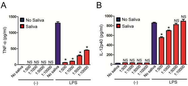
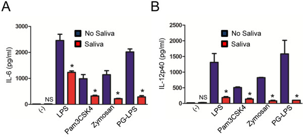
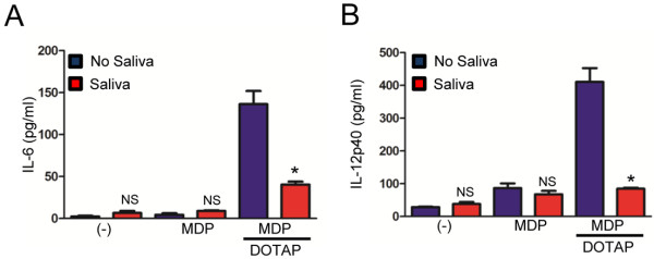
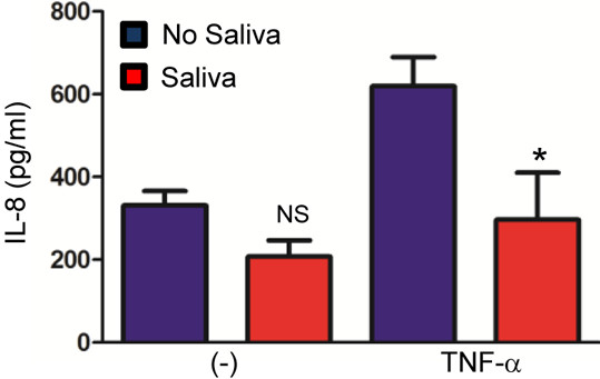
Methods: Bone marrow derived macrophages (BMDMs) were stimulated with pathogen associated molecular patterns (PAMPs) and A. phagocytophilum. Cytokine secretion was measured in the presence and absence of I. scapularis saliva. Human peripheral blood mononuclear cells (PBMCs) were also stimulated with Tumor Necrosis Factor (TNF)-α in the presence and absence of I. scapularis saliva and interleukin (IL)-8 was measured.

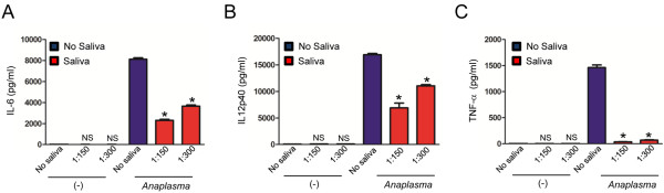
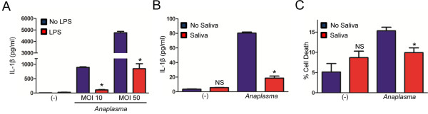
Results: I. scapularis saliva inhibits inflammatory cytokine secretion by macrophages during stimulation of Toll-like (TLR) and Nod-like receptor (NLR) signaling pathways. The effect of I. scapularis saliva on immune cells is not restricted to murine macrophages because decreasing levels of interleukin (IL)-8 were observed after TNF-α stimulation of human peripheral blood mononuclear cells. I. scapularis saliva also mitigates pro-inflammatory cytokine response by murine macrophages during challenge with A. phagocytophilum.

Conclusions: These findings suggest that I. scapularis may inhibit inflammatory cytokine secretion during rickettsial transmission at the vector-host interface.


Figures

Figure 1
Figure 1
I. scapularis saliva mitigates LPS-mediated cytokine secretion by murine macrophages. BMDMs (×ばつ105) from C57BL/6 mice were stimulated with LPS (500 ng/ml) for 18 hours, in the presence or absence of indicated dilutions of tick saliva. (A) TNF-α and (B) IL-12p40 were measured by ELISA. Tick saliva was added 2 hours before stimulation. Responses were measured in triplicate and presented as mean ± SEM within the representative experiment. Experiments were repeated three times. *P < .05, One-way ANOVA, post-hoc Bonferroni; (−) non-stimulated cells. NS – not significant.
Figure 2
Figure 2
I. scapularis saliva inhibits TLR-mediated cytokine secretion by macrophages. BMDMs (×ばつ105) from C57BL/6 mice were stimulated with LPS (500 ng/ml), Pam3CSK4 (1 μg/ml), Zymosan (10 μg/ml) and PG-LPS (500 ng/ml) for 18 hours in the presence or absence of tick saliva (1:500 dilution). Tick saliva was added 2 hours before stimulation. (A) IL-6 and (B) IL-12p40 were measured by ELISA. Responses were measured in triplicate and presented as mean ± SEM within the representative experiment. Experiments were repeated three times. *P < .05, Student’s t test. (−) non-stimulated cells. NS – not significant.
Figure 3
Figure 3
I. scapularis saliva impairs Nod2-mediated cytokine secretion by murine macrophages. BMDMs (×ばつ106) from C57BL/6 mice were stimulated with MDP (10 μg/ml) or DOTAP (10 μg/ml) + MDP (10 μg/ml) for 20 hours, in the presence or absence of tick saliva (1:500). The secretion of (A) IL-6 and (B) IL-12p40 were measured by ELISA. Responses were measured in triplicate and presented as mean ± SEM within the representative experiment. Experiments were repeated three times. *P < .05, Student’s t test. (−) non-stimulated cells. NS – not significant.
Figure 4
Figure 4
I. scapularis saliva decreases IL-8 secretion by human peripheral blood mononuclear cells. PBMCs (×ばつ105) were purified and plated for 2 hours. Cells were incubated with tick saliva for 30 minutes followed by 4 hours of stimulation with 100 ng/ml TNF-α. The secretion of IL-8 (CXCL8) was measured by ELISA. Responses were measured in triplicate and presented as mean ± SEM within the representative experiment. *P < .05, Student’s t test. (−) non-stimulated cells. NS – not significant.
Figure 5
Figure 5
I. scapularis saliva mitigates cytokine secretion by macrophages during A. phagocytophilum stimulation in a dose-dependent manner. BMDMs (×ばつ106) from C57BL/6 mice were stimulated with the wild-type A. phagocytophilum HZ strain (MOI 50) for 18 hours in the presence or absence of tick saliva (1:150 and 1:300 dilution). (A) IL-6, (B) IL-12p40 and (C) TNF-α were measured by ELISA. Responses were measured in triplicate and presented as mean ± SEM within the representative experiment. *P < .05, One-way ANOVA, post-hoc Bonferroni; (-) non-stimulated cells. NS – not significant.
Figure 6
Figure 6
I. scapularis saliva decreases IL-1β secretion by macrophages during A. phagocytophilum stimulation. (A) BMDMs (×ばつ106) from C57BL/6 mice were primed with LPS (500 ng/nl) for 24 hours, washed and then stimulated with the A. phagocytophilum HZ strain (MOIs 10 and 50) for another 24 hours. IL-1β was measured by ELISA. (B) BMDMs (×ばつ106) from C57BL/6 mice were stimulated with the wild-type A. phagocytophilum HZ strain (MOI 50) for 18 hours in the presence or absence of tick saliva (1:150 dilution). IL-1β was measured by ELISA. (C) Cell death was measured using the LDH assay. Responses were measured in triplicate and presented as mean ± SEM within the representative experiment. *P < .05, Student’s t test. (−) non-stimulated cells. NS – not significant.

References

    1. Francischetti IM, Sa-Nunes A, Mans BJ, Santos IM, Ribeiro JM. The role of saliva in tick feeding. Front Biosci. 2009;14:2051–2088. - PMC - PubMed
    1. Fontaine A, Diouf I, Bakkali N, Misse D, Pages F, Fusai T, Rogier C, Almeras L. Implication of haematophagous arthropod salivary proteins in host-vector interactions. Parasit Vectors. 2011;4:187. doi: 10.1186/1756-3305年4月18日7. - DOI - PMC - PubMed
    1. Andersen JF. Structure and mechanism in salivary proteins from blood-feeding arthropods. Toxicon. 2010;56(7):1120–1129. doi: 10.1016/j.toxicon.200911002. - DOI - PMC - PubMed
    1. Chmelar J, Calvo E, Pedra JH, Francischetti IM, Kotsyfakis M. Tick salivary secretion as a source of antihemostatics. J Proteomics. 2012;75(13):3842–3854. doi: 10.1016/j.jprot.2012年04月02日6. - DOI - PMC - PubMed
    1. Valenzuela JG. Exploring tick saliva: from biochemistry to ‘sialomes’ and functional genomics. Parasitology. 2004;129(Suppl):S83–S94. - PubMed

Publication types

Cite

AltStyle によって変換されたページ (->オリジナル) /