This site needs JavaScript to work properly. Please enable it to take advantage of the complete set of features!
Skip to main page content
U.S. flag

An official website of the United States government

Dot gov

The .gov means it’s official.
Federal government websites often end in .gov or .mil. Before sharing sensitive information, make sure you’re on a federal government site.

Https

The site is secure.
The https:// ensures that you are connecting to the official website and that any information you provide is encrypted and transmitted securely.

NIH NLM Logo
Log in
Access keys NCBI Homepage MyNCBI Homepage Main Content Main Navigation
Review
. 2012 Oct;28(10):395-407.
doi: 10.1016/j.pt.201207006. Epub 2012 Sep 1.

The tumorigenic liver fluke Opisthorchis viverrini--multiple pathways to cancer

Affiliations
Review

The tumorigenic liver fluke Opisthorchis viverrini--multiple pathways to cancer

Banchob Sripa et al. Trends Parasitol. 2012 Oct.

Abstract

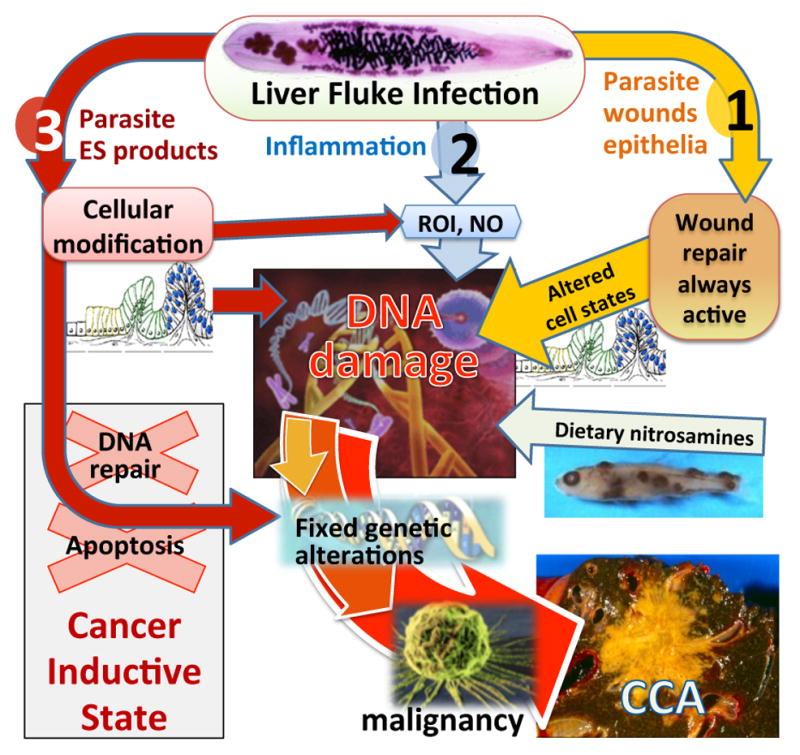
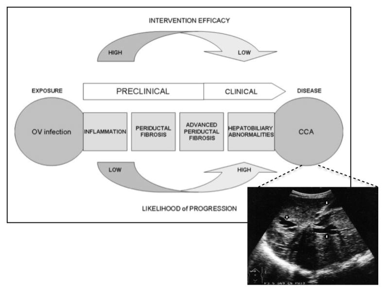
Liver fluke infection caused by Opisthorchis viverrini is a major public health problem in Thailand and adjacent countries. In addition to infection-associated morbidity, infection with O. viverrini and the related Clonorchis sinensis are unarguable risk factors for cholangiocarcinoma (CAA, bile-duct cancer). Here we review the pathogenesis of opisthorchiasis and the association between O. viverrini infection and bile-duct cancer, focusing on the molecular parallels between wound healing, chronic inflammation, and cancer development. We review a schema for human disease progression from fluke infection, chronic opisthorchiasis, advanced periductal fibrosis, and cholangiocarcinogenesis, and present a rationale for biomarker discovery to facilitate early intervention. We conclude by addressing post-genomic advances with a view to developing new control strategies to combat this infectious cancer.


Figures

Figure 1
Figure 1. Incidence of liver cancer and prevalence of liver fluke infections
Liver cancer rates are divided into the major subtypes of hepatocellular carcinoma (HCC), cholangiocarcinoma (CCA) and other less common subtypes (a). Regions within Thailand are highlighted with red stars. The prevalence of O. viverrini and C. sinensis in the Mekong Basin sub-region (b). Endemicity level is defined based on prevalence of infections: low – 1–5%; medium - 5.1–15%; high - >15%. Taken from [6].
Figure 2
Figure 2. Hypothesized pathways of pathogenesis of liver fluke-induced cholangiocarcinoma (CCA)
O. viverrini damages bile duct epithelia via three pathways: (1) mechanical damage by feeding parasites; (2) immunopathology, particularly due to reactive oxygen intermediates (ROI) and nitric oxide (NO); (3) direct effects of fluke secreted proteins including induction of cell proliferation and inhibiting cancer prevention pathways (DNA repair/apoptosis). These pathways converge, resulting in genetic lesions that become fixed after successive replications, eventually leading to malignant transformation of cholangiocytes into CCA. Adapted from [121].
Figure 3
Figure 3. Immunolocalization of Ov-GRN-1 in adult O. viverrini and bile ducts of experimentally infected hamsters
The left panel was probed with IgG purified from normal mouse serum (NMS); the right panel was probed with anti-Ov-GRN-1 IgG. Sections were stained with immunoperoxidase revealed as a brown/rust colored deposit and Mayer s Haematoxylin counterstained the nuclei in blue. Red arrows highlight the regions within the O. viverrini parasite and bile duct tissue that stained positive for Ov-GRN-1. From [41].
Figure 4
Figure 4. Epithelial hyperplasia and DNA damage in the bile ducts of O. viverrini infected hamsters
(a) Proliferating epithelial cells were detected in the bile ducts surrounding adult flukes (Ov) using bromodeoxyuridine (arrow); ×ばつ200 magnification. (b) DNA damage in epithelial cells (arrow) revealed by 8-oxodG staining in the liver of hamsters that were chronically infected with O. viverrini (Ov). Normal liver did not stain positive for either stain (not shown). Photo provided by B. Sripa (unpublished).
Figure 5
Figure 5. Conceptual framework for O. viverrini-induced cholangiocarcinoma (CCA)
The primary exposure (OV infection) is identified and the progression of disease events to CCA are tracked in well-defined stages. Linked to the CCA window of the schematic is an ultrasonograph depicting a CCA. Note liver mass surrounded by dilated intrahepatic ducts (arrows). Image provided by EM. Figure adapted and modified for O. viverrini-induced CCA from [27].
Figure 6
Figure 6. Plasma IL-6 concentrations for detection of O. viverrini-induced advanced periductal fibrosis (left panel) or cholangiocarcinoma (right panel)
A receiver-operating-characteristic (ROC) curve plots the True Positive Probability (sensitivity) against the False Negative Probability (1 – specificity) for the full range of IL-6 cut-off points for the detection of O. viverrini-induced APF or CCA. The area under the ROC curve is interpreted as the probability of correctly identifying (accuracy) a randomly selected participant as either a case (APF positive or CCA) or a non-case (APF negative or CCA negative). The 45-degree line in the graph subsumes an area equal to 0.50 (50%), which is equivalent to using a coin toss procedure to classify participants as either cases or controls. Figure adapted from [27].
Figure 7
Figure 7. Effect of praziquantel on expression of inducible nitric oxide synthase (iNOS) and nuclear factor- κB (NF-κB) in infected hamsters
Expression of iNOS and NF-κB in hamster livers was assessed by double immunofluorescence. O. viverrini infection induced iNOS expression (red) in the cytoplasm and NF-κB accumulation (green) in the nucleus of bile duct epithelial cells. Treatment with praziquantel gradually increased expression of these proteins 6 hours post-treatment. Expression reached its highest level 12 hours post-treatment and then decreased 24 hours post-treatment. Normal hamsters treated with praziquantel and analyzed 12 hours after treatment (N + PZ, 12 hr) showed weak immunoreactivity after prolonged exposure (microscopy) to highlight minimal background fluorescence. Magnification ×ばつ 400; magnification ×ばつ 200 for N + PZ. OV - O. viverrini; Bd - bile duct. Taken from [74].

References

    1. Lun ZR, et al. Clonorchiasis: a key foodborne zoonosis in China. Lancet Infect Dis. 2005;5:31–41. - PubMed
    1. Hong ST, Fang Y. Clonorchis sinensis and clonorchiasis, an update. Parasitol Int. 2012;61:17–24. - PubMed
    1. Keiser J, Utzinger J. Food-borne trematodiases. Clin Microbiol Rev. 2009;22:466–483. - PMC - PubMed
    1. Mordvinov VA, et al. Opisthorchis felineus and Metorchis bilis are the main agents of liver fluke infection of humans in Russia. Parasitol Int. 2012;61:25–31. - PubMed
    1. IARC. Schistosomes, liver flukes and Helicobacter pylori. IARC Working Group on the Evaluation of Carcinogenic Risks to Humans. Lyon, 7–14 June 1994. IARC Monogr Eval Carcinog Risks Hum. 1994;61:1–241. - PMC - PubMed

Publication types

Cite

AltStyle によって変換されたページ (->オリジナル) /