This site needs JavaScript to work properly. Please enable it to take advantage of the complete set of features!
Skip to main page content
U.S. flag

An official website of the United States government

Dot gov

The .gov means it’s official.
Federal government websites often end in .gov or .mil. Before sharing sensitive information, make sure you’re on a federal government site.

Https

The site is secure.
The https:// ensures that you are connecting to the official website and that any information you provide is encrypted and transmitted securely.

NIH NLM Logo
Log in
Access keys NCBI Homepage MyNCBI Homepage Main Content Main Navigation
doi: 10.1371/journal.pone.0043207. Epub 2012 Aug 15.

Development and characterization of a caprine aerosol infection model of melioidosis

Affiliations

Development and characterization of a caprine aerosol infection model of melioidosis

Carl Soffler et al. PLoS One. 2012.

Abstract

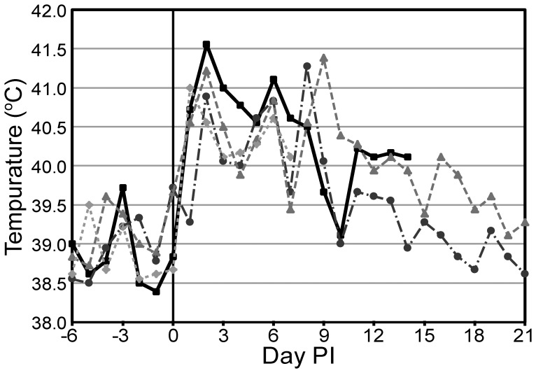
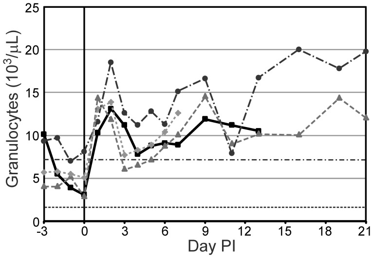
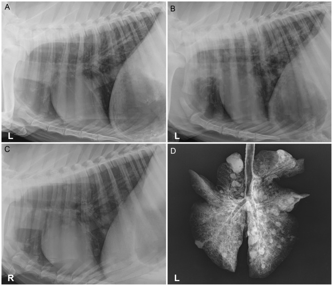
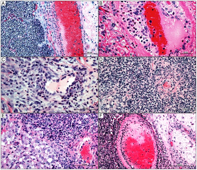
Infection with Burkholderia pseudomallei causes the disease melioidosis, which often presents as a serious suppurative infection that is typically fatal without intensive treatment and is a significant emerging infectious disease in Southeast Asia. Despite intensive research there is still much that remains unknown about melioidosis pathogenesis. New animal models of melioidosis are needed to examine novel aspects of pathogenesis as well as for the evaluation of novel therapeutics. The objective of the work presented here was to develop a subacute to chronic caprine model of melioidosis and to characterize the progression of disease with respect to clinical presentation, hematology, clinical microbiology, thoracic radiography, and gross and microscopic pathology. Disease was produced in all animals following an intratracheal aerosol of 10(4) CFU delivered, with variable clinical manifestations indicative of subacute and chronic disease. Bronchointerstitial pneumonia was apparent microscopically by day 2 and radiographically and grossly apparent by day 7 post infection (PI). Early lesions of bronchopneumonia soon progressed to more severe bronchointerstitial pneumonia with pyogranuloma formation. Extrapulmonary dissemination appeared to be a function of pyogranuloma invasion of pulmonary vasculature, which peaked around day 7 PI. Histopathology indicated that leukocytoclastic vasculitis was the central step in dissemination of B. pseudomallei from the lungs as well as in the establishment of new lesions. While higher doses of organism in goats can produce acute fatal disease, the dose investigated and resulting disease had many similarities to human melioidosis and may warrant further development to provide a model for the study of both natural and bioterrorism associated disease.


Conflict of interest statement

Competing Interests: The authors have declared that no competing interests exist.

Figures

Figure 1
Figure 1. Temperatures from selected goats.
Peaks in temperature are seen on days 1 and 2 and between days 6 and 9. Based on the temporospatial distribution of gross lesions, these peaks are likely associated with bacteremic dissemination events even though bacteremia was not detected with blood culture. BpG15 (square), BpG21 (circle), BpG22 (triangle), BpG27 (diamond).
Figure 2
Figure 2. Granulocyte counts from selected goats.
During the first half of the study, the granulocyte count appears to mirror the temperature trends seen in Figure 1. BpG15 (square), BpG21 (circle), BpG22 (triangle), BpG27 (diamond), RR High (dash-dot)/Low (dot-dot): reference range limits of normal caprine granulocyte count.
Figure 3
Figure 3. Thoracic and extirpated lung radiographs of goats infected with an intratracheal aerosol of
A) Goat 15, day 7, left lateral thorax with a mild bronchointerstitial pattern restricted to the caudal lung lobes. B) Goat 16, day 7, left lateral thorax with moderate to severe bronchointerstitial infiltrates with numerous ill-defined variably sized nodules in all lung lobes with the caudal lung lobes more affected. C) Goat 22, day 21, right lateral thorax with small nodules seen in all lung lobes and mild-moderate bronchointerstitial infiltrates diffusely. D) Goat 22, day 21, extirpated lungs, numerous nodules in all lung lobes, which are much more distinct than in the in vivo radiographs.
Figure 4
Figure 4. Gross and histologic lesions of caprine melioidosis.
A) Lung, multiple discrete subpleural targetoid pyogranulomas with tan purulent centers and consolidated hyperemic rims; B) a splenic subcapsular pyogranuloma bulging over the splenic surface with regional capsulitis and injected vessels; C) bronchiolar epithelium showing apical surface deciliation, segmental necrosis of the pseudostratified ciliated epithelium, neutrophil transcytosis, and luminal aggregates of neutrophils suspended in inflammatory edema; D) pulmonary septal thickening secondary to neutrophil infiltration; E) sever necrosuppurative/ulcerative tracheitis with neutrophil transcytosis and a mucosal abscess (inset); F) linear renal pyogranuloma extending from the cortex down into the medulla obliterating large areas of renal parenchyma. 4C–4F hematoxylin and eosin staining.
Figure 5
Figure 5. Caprine melioidosis vasculitides in multiple organs.
A) pulmonary pyogranuloma encroaching on large interlobular vein, resulting in a segmental leukocytoclastic vasculitis – unaffected endothelium is present on the contralateral side of the vessel; B) pulmonary vasculitis in a medium-sized artery demonstrating a clear separation of the tunica intima from the tunica media by neutrophils and macrophages with segmental intimal proliferation; C) splenic capsular vasculitis with perivascular neutrophilic infiltrate; D) vasocentric renal pyogranuloma showing neutrophil infiltration into the subintima and adventitia with endothelial proliferation and fibrinoid necrosis of a small renal vessel E) focal vasculitis and early pyogranuloma formation at the corticomedullary junction of the adrenal gland; F) leukocytoclastic arteritis in the testicle with an adjacent area of testicular degeneration showing diminished stratification of spermatogenic cells and interstitial suppurative orchitis. 5A–5F, hematoxylin and eosin staining.

References

    1. Whitmore A, Krishnaswami CS (1912) An account of the discovery of a hitherto underscribed infective disease occurring among the population of Rangoon. The Indian Medical Gazette 47: 262–267. - PMC - PubMed
    1. Currie BJ, Dance DAB, Cheng AC (2008) The global distribution of Burkholderia pseudomallei and melioidosis: an update. Transactions of the Royal Society of Tropical Medicine and Hygiene 102: S1–S4. - PubMed
    1. Cheng AC, Currie BJ (2005) Melioidosis: epidemiology, pathophysiology, and management. Clin Microbiol Rev 18: 383–416. - PMC - PubMed
    1. Ngauy V, Lemeshev Y, Sadkowski L, Crawford G (2005) Cutaneous melioidosis in a man who was taken as a prisoner of war by the Japanese during World War II. J Clin Microbiol 43: 970–972. - PMC - PubMed
    1. Gilad J, Harary I, Dushnitsky T, Schwartz D, Amsalem Y (2007) Burkholderia mallei and Burkholderia pseudomallei as bioterrorism agents: national aspects of emergency preparedness. Isr Med Assoc J 9: 499–503. - PubMed

Publication types

Cite

AltStyle によって変換されたページ (->オリジナル) /