Immunobiology of visceral leishmaniasis
- PMID: 22912637
- PMCID: PMC3418610
- DOI: 10.3389/fimmu.2012.00251
Immunobiology of visceral leishmaniasis
Abstract
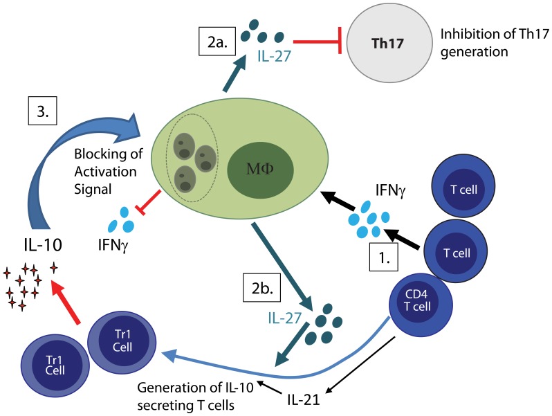
Visceral leishmaniasis (VL), commonly known as kala-azar, is caused by Leishmania donovani and Leishmania infantum (Leishmania chagasi in the Americas). These Leishmania species infect macrophages throughout the viscera, and parasites are typically found in the spleen, liver, and bone marrow. Patients with active disease typically exhibit marked immunosuppression, lack reactivity to the Leishmania skin test (LST), a delayed type hypersensitivity test, and their peripheral blood mononuclear cells (PBMC) fail to respond when stimulated with leishmanial antigens in vitro. However, most people infected with visceralizing species of Leishmania never develop disease. Understanding immune failure and the underlying immune mechanism that lead to disease as well as control of infection are key questions for research in this field. In this review, we discuss immunological events described in human and experimental VL and how these can affect the outcome of infection.
Keywords: IL-10; Leishmania donovani; T cells; immune regulation; visceral leishmaniasis.
Figures
References
-
- Agrawal S., Rai M., Sundar S. (2005). Management of visceral leishmaniasis: Indian perspective. J. Postgrad. Med. 51(Suppl. 1) S53–S57 - PubMed
-
- Alvar J., Canavate C., Molina R., Moreno J., Nieto J. (2004). Canine leishmaniasis. Adv. Parasitol. 57 1–88 - PubMed
-
- Anam K., Afrin F., Banerjee D., Pramanik N., Guha S. K., Goswami R. P., Gupta P. N., Saha S. K., Ali N. (1999). Immunoglobulin subclass distribution and diagnostic value of Leishmania donovani antigen-specific immunoglobulin G3 in Indian kala-azar patients. Clin. Diagn. Lab. Immunol. 6 231–235 - PMC - PubMed
-
- Anderson C. F., Lira R., Kamhawi S., Belkaid Y., Wynn T. A., Sacks D. (2008). IL-10 and TGF-beta control the establishment of persistent and transmissible infections produced by Leishmania tropica in C57BL/6 mice. J. Immunol. 180 4090–4097 - PubMed
LinkOut - more resources
Full Text Sources