This site needs JavaScript to work properly. Please enable it to take advantage of the complete set of features!
Skip to main page content
U.S. flag

An official website of the United States government

Dot gov

The .gov means it’s official.
Federal government websites often end in .gov or .mil. Before sharing sensitive information, make sure you’re on a federal government site.

Https

The site is secure.
The https:// ensures that you are connecting to the official website and that any information you provide is encrypted and transmitted securely.

NIH NLM Logo
Log in
Access keys NCBI Homepage MyNCBI Homepage Main Content Main Navigation
. 2012;6(8):e1789.
doi: 10.1371/journal.pntd.0001789. Epub 2012 Aug 14.

Phenotypic characterization of peripheral T cells and their dynamics in scrub typhus patients

Affiliations

Phenotypic characterization of peripheral T cells and their dynamics in scrub typhus patients

Bon-A Cho et al. PLoS Negl Trop Dis. 2012.

Abstract

Background: Scrub typhus, caused by Orientia tsutsugamushi infection, is one of the main causes of febrile illness in the Asia-Pacific region. Although cell-mediated immunity plays an important role in protection, little is known about the phenotypic changes and dynamics of leukocytes in scrub typhus patients.

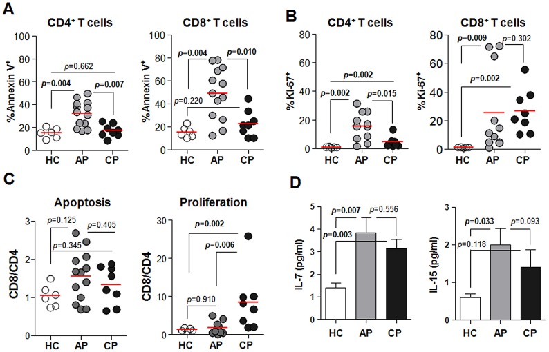
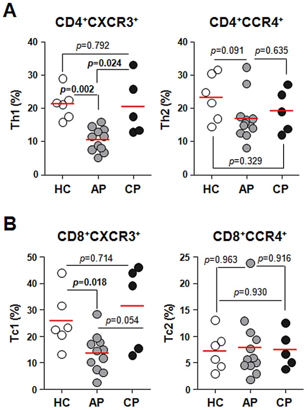
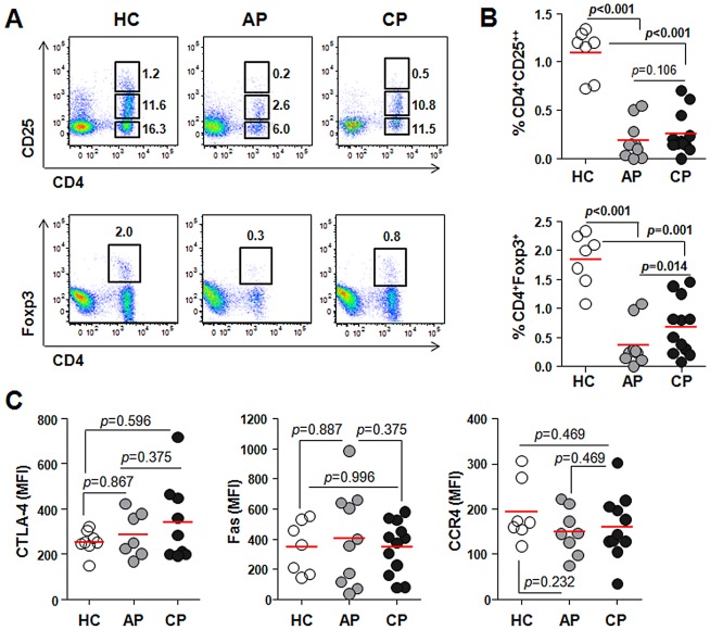
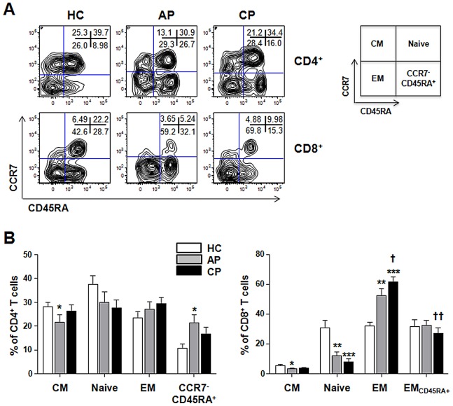
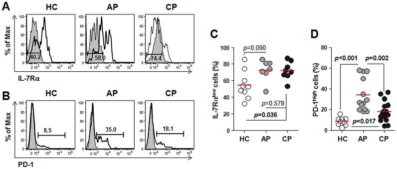
Methodology/principal findings: To reveal the underlying mechanisms of immunological pathogenesis, we extensively analyzed peripheral blood leukocytes, especially T cells, during acute and convalescent phases of infection in human patients and compared with healthy volunteers. We observed neutrophilia and CD4(+) T lymphopenia in the acute phase of infection, followed by proliferation of CD8(+) T cells during the convalescent phase. Massive T cell apoptosis was detected in the acute phase and preferential increase of CD8(+) T cells with activated phenotypes was observed in both acute and convalescent phases, which might be associated or correlated with elevated serum IL-7 and IL-15. Interestingly, peripheral Treg cells were significantly down-regulated throughout the disease course.

Conclusions/significance: The remarkable decrease of CD4(+) T cells, including Treg cells, during the acute phase of infection may contribute to the loss of immunological memory that are often observed in vaccine studies and recurrent human infection.


Conflict of interest statement

The authors have declared that no competing interests exist.

Figures

Figure 1
Figure 1. Profiles of peripheral blood leukocytes and T lymphocytes in scrub typhus patients.
A. The frequencies (%) of neutrophils, lymphocytes, and monocytes in blood leukocytes from healthy controls (HC, n = 9, open circle) were determined using a hematology analyzer and compared to frequencies in scrub typhus patients during acute phase (AP, n = 17, gray circle) or convalescent phase (CP, n = 17, black circle) of infection. B. Peripheral blood mononuclear cells (PBMCs) from scrub typhus patients and healthy controls were stained with anti-CD3 and CD8 antibodies and analyzed by flow cytometry (see Figure S1). The frequencies (% in lymphocytes) and absolute numbers (cells/mm2) of CD4+ or CD8+ T cells in PBMCs of the patients (AP, n = 15 and CP, n = 15) and healthy controls (HC, n = 9) were compared. The absolute number of each leukocyte subset was calculated based on the leukocyte differential counts and the frequency information obtained from flow cytometry. For example, CD4+ T cells count = (lymphocyte count) x (% of CD4+ T cells)/100. The data of each individual person are presented in Table S3. Red bars indicate the mean value and p values were obtained using the Mann-Whitney U test or Wilcoxon signed-rank test. Statistically significant p values (<0.05) are shown in bold.
Figure 2
Figure 2. Apoptosis and proliferation of T cells during Orientia infection.
A and B. PBMCs were stained with antibodies against CD4, CD8, Annexin V, or Ki-67 and then analyzed by flow cytometry. The frequencies of Annexin V- (A) or Ki-67- (B) positive cells in CD4+ and CD8+ T cells were compared among healthy controls (HC, n = 6, open circle) and scrub typhus patients at acute phase (AP, n = 13–15, gray circle) or convalescent phase (CP, n = 6, black circle). Red bars indicate the mean value. C. The CD8/CD4 ratio of apoptotic or proliferating cells were compared among the patients and healthy volunteers. D. The levels of IL-7 and IL-15 (pg/ml) in the sera were measured and compared. Error bars indicate standard error from the mean value (C and D). p values were obtained using the Mann-Whitney U test. Statistically significant p values (<0.05) are shown in bold.
Figure 3
Figure 3. Profiles of type 1 and type 2 T cells in peripheral blood of scrub typhus patients.
PBMCs were stained with antibodies against CXCR3, CCR4, CD4, and CD8 and then analyzed on a flow cytometer. A. The frequencies of Th1 (CD4+CXCR3+) or Th2 (CD4+CCR4+) within CD4+ T cells were compared among healthy controls (HC, n = 6, open circle) and the patients in acute phase (AP, n = 12, gray circle) or convalescent phase (CP, n = 5, black circle). B. The frequencies of Tc1 (CD8+CXCR3+) and Tc2 (CD8+CCR4+) within CD8+ T cells were compared among healthy controls and the patients as in A. Red bars indicate the mean value and p values were obtained using the Mann-Whitney U test. Statistically significant p values (<0.05) are shown in bold.
Figure 4
Figure 4. Profiles of CD4+Foxp3+ or CD4+CD25++ regulatory T cells in scrub typhus patients.
A. PBMCs were stained with antibodies against CD4 and CD25 or Foxp3 and then analyzed on a flow cytometer. Representative dot plots show the identification of CD25++ T cells (upper panels) or Foxp3+ T cells (lower panels) within CD4+ T cells. Numbers in the plots indicate the frequencies (%) of the gated cells in total PBMCs. B. The frequency of CD4+CD25++ or CD4+Foxp3+ T cells were compared between healthy controls (HC, n = 7, open circle) and scrub typhus patients at acute phase (AP, n = 10, gray circle) or convalescent phase (CP, n = 12, black circle). C. PBMCs were stained with antibodies against CD4 and Fas or CCR4, followed by intracellular staining of Foxp3 and/or CTLA-4 after fixation and permeablization. Mean fluorescent Intensities (MFI) representing CTLA-4, Fas, or CCR4 expression in CD4+Foxp3+ regulatory cells from healthy controls (HC, n = 7–8, open circle) or the patients (AP, n = 7–10, gray circle and CP, n = 9–12, black circle) were compared. Red bars indicate the mean value and p values were obtained using the Mann-Whitney U test or Wilcoxon signed-rank test. Statistically significant p values (<0.05) are shown in bold.
Figure 5
Figure 5. Changes of effector and memory T cell subsets in scrub typhus patients.
A. Representative contour plots show the frequencies of each subsets within CD4+ (upper panels) or CD8+ (lower panels) T cell populations. Numbers within the plots indicate the percentage of each subset. B. The frequencies of naïve (CCR7+CD45RA+), central memory (CM, CCR7+CD45RA), and effector memory (EM, CCR7CD45RA), CD45RA+ EM (EMCD45RA+, for CD8+ T cells), or CCR7CD45RA+ (for CD4+ T cells) T-cell subsets in healthy controls (HC, n = 9) and scrub typhus patients at acute phase (AP, n = 15) or convalescent phase (CP, n = 17) were compared. Error bars indicate standard error of mean values. p values were obtained using the Mann-Whitney U test or Wilcoxon signed-rank test. *, p<0.05; **, p<0.01; and ***, p<0.001 (compared with that of healthy control); †, p<0.05 and ††, p<0.01 (compared with that of the patients at acute phase).
Figure 6
Figure 6. Analysis of activated CD8+ T cells in peripheral bloods of scrub typhus patients.
PBMCs were stained with antibodies against CD8 and PD-1 or IL-7Rα and then analyzed on a flow cytometer. A and B. Representative histograms show the frequencies of IL-7Rαlow cells (A) and PD-1high cells (B) within CD8+ T cells. Gray histogram, isotype control. C. The frequency of IL-7RαlowCD8+ T cells from healthy controls (HC, n = 9, open circle) is compared with that of scrub typhus patients at acute phase (AP, n = 7, gray circle) or convalescent phase (CP, n = 8, black circle). D. The frequencies of PD-1highCD8+ T cells were compared among healthy controls (n = 9) and the patients (AP, n = 13; CP, n = 15). Red bars indicate the mean value and p values were obtained using the Mann-Whitney U test or Wilcoxon signed-rank test. Statistically significant p values (<0.05) are shown in bold.

References

    1. Seong SY, Choi MS, Kim IS (2001) Orientia tsutsugamushi infection: overview and immune responses. Microbes Infect 3: 11–21. - PubMed
    1. Richards AL (2004) Rickettsial vaccines: the old and the new. Expert Rev Vaccines 3: 541–555. - PubMed
    1. Kweon SS, Choi JS, Lim HS, Kim JR, Kim KY, et al. (2009) Rapid increase of scrub typhus, South Korea, 2001–2006. Emerg Infect Dis 15: 1127–1129. - PMC - PubMed
    1. Mathai E, Rolain JM, Verghese GM, Abraham OC, Mathai D, et al. (2003) Outbreak of scrub typhus in southern India during the cooler months. Ann N Y Acad Sci 990: 359–364. - PubMed
    1. Zhang S, Song H, Liu Y, Li Q, Wang Y, et al. (2010) Scrub typhus in previously unrecognized areas of endemicity in China. J Clin Microbiol 48: 1241–1244. - PMC - PubMed

Publication types

Cite

AltStyle によって変換されたページ (->オリジナル) /