This site needs JavaScript to work properly. Please enable it to take advantage of the complete set of features!
Skip to main page content
U.S. flag

An official website of the United States government

Dot gov

The .gov means it’s official.
Federal government websites often end in .gov or .mil. Before sharing sensitive information, make sure you’re on a federal government site.

Https

The site is secure.
The https:// ensures that you are connecting to the official website and that any information you provide is encrypted and transmitted securely.

NIH NLM Logo
Log in
Access keys NCBI Homepage MyNCBI Homepage Main Content Main Navigation
. 2012 Jun 27:5:109.
doi: 10.1186/1756-3305年5月10日9.

The life cycle of Trypanosoma (Nannomonas) congolense in the tsetse fly

Affiliations

The life cycle of Trypanosoma (Nannomonas) congolense in the tsetse fly

Lori Peacock et al. Parasit Vectors. .

Abstract

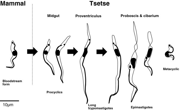
Background: The tsetse-transmitted African trypanosomes cause diseases of importance to the health of both humans and livestock. The life cycles of these trypanosomes in the fly were described in the last century, but comparatively few details are available for Trypanosoma (Nannomonas) congolense, despite the fact that it is probably the most prevalent and widespread pathogenic species for livestock in tropical Africa. When the fly takes up bloodstream form trypanosomes, the initial establishment of midgut infection and invasion of the proventriculus is much the same in T. congolense and T. brucei. However, the developmental pathways subsequently diverge, with production of infective metacyclics in the proboscis for T. congolense and in the salivary glands for T. brucei. Whereas events during migration from the proventriculus are understood for T. brucei, knowledge of the corresponding developmental pathway in T. congolense is rudimentary. The recent publication of the genome sequence makes it timely to re-investigate the life cycle of T. congolense.

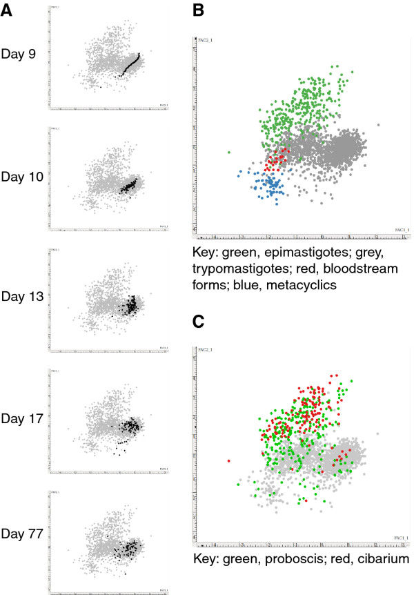
Methods: Experimental tsetse flies were fed an initial bloodmeal containing T. congolense strain 1/148 and dissected 2 to 78 days later. Trypanosomes recovered from the midgut, proventriculus, proboscis and cibarium were fixed and stained for digital image analysis. Trypanosomes contained in spit samples from individually caged flies were analysed similarly. Mensural data from individual trypanosomes were subjected to principal components analysis.

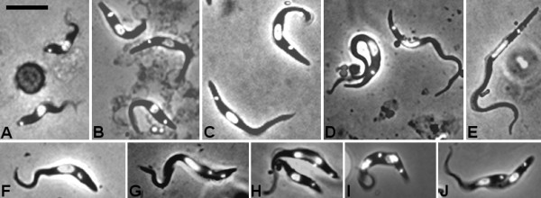
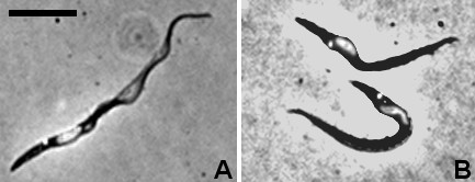
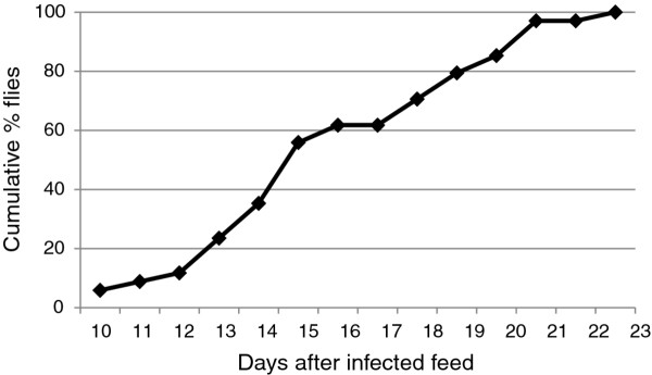
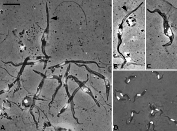
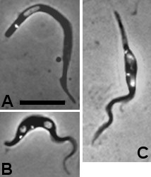
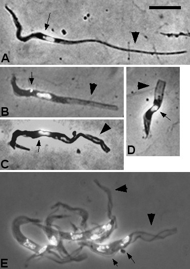
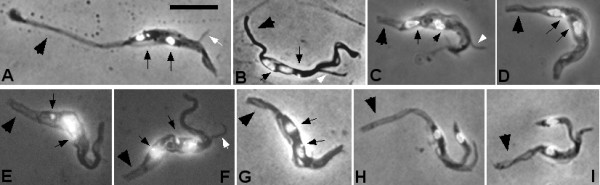
Results: Flies were more susceptible to infection with T. congolense than T. brucei; a high proportion of flies infected with T. congolense established a midgut and subsequent proboscis infection, whereas many T. brucei infections were lost in the migration from foregut to salivary glands. In T. congolense, trypomastigotes ceased division in the proventriculus and became uniform in size. The trypanosomes retained trypomastigote morphology during migration via the foregut to the mouthparts and we confirmed that the trypomastigote-epimastigote transition occurred in the proboscis. We found no equivalent to the asymmetric division stage in T. brucei that mediates transition of proventricular trypomastigotes to epimastigotes. In T. congolense extremely long epimastigotes with remarkably elongated posterior ends were observed in both the proboscis and cibarium; no difference was found in the developmental stages in these two organs. Dividing trypomastigotes and epimastigotes were recovered from the proboscis, some of which were in transition from trypomastigote to epimastigote and vice versa. It remains uncertain whether these morphological transitions are mediated by cell division, since we also found non-dividing cells with a variously positioned, juxta-nuclear kinetoplast.

Conclusions: We have presented a detailed description of the life cycle of T. congolense in its tsetse fly vector. During development in the fly T. congolense shares a common migratory pathway with its close relative T. brucei, culminating in the production of small metacyclic trypanosomes that can be inoculated with the saliva. Despite this outward similarity in life cycle, the transitional developmental stages in the foregut and mouthparts are remarkably different in the two trypanosome species.


Figures

Figure 1
Figure 1
Morphology of developmental stages ofTrypanosoma congolense. Fixed and DAPI-stained cells; each panel shows a merge of DAPI and brightfield images. A. Bloodstream forms. Panels B to F are procyclic trypomastigotes from the tsetse midgut 2 days (B), 6 days (C), 9 days (D) and 17 days (E) after ingestion of the infected bloodmeal. Panels F to J are trypomastigotes from the tsetse midgut in various stages of division; 2K1N (F, G), 2K2N (H-J). Bar = 10 μm.
Figure 2
Figure 2
Proventricular trypanosomes. A. Trypomastigote with posterior kinetoplast. B. Trypomastigotes with the kinetoplast adjacent and to the posterior pole of the nucleus; the cell is distended in the region of the kinetoplast and nucleus. Bar = 10 μm.
Figure 3
Figure 3
Principal components analysis (PCA). Each plot shows scores for PCA factor 1 versus factor 2 derived from the mensural data from 2205 individual trypanosomes, each represented by a coloured dot. A. Sequential plots of proventricular trypanosomes (black dots) compared with all other trypanosomes (grey dots). B. Comparison of bloodstream forms (red dots), midgut, proboscis and cibarium trypomastigotes (grey dots), epimastigotes (green dots), and metacyclics (blue dots). C. Comparison of trypanosomes from the proboscis and cibarium.
Figure 4
Figure 4
Trypanosoma congolensein spit samples. Cumulative percentage of infected flies that produced a trypanosome-positive spit sample. A total of 34 flies produced a trypanosome-positive sample during the timecourse of 10–23 days.
Figure 5
Figure 5
Trypanosomes in spit samples 16–21 days after infection. A. Mixture of trypomastigotes with epimastigote (arrowed). B. Epimastigote with long posterior (arrowed). C. Epimastigote (2K1N). D. Metacyclics from day 21; contrast the size of metacyclics with the other trypomastigotes from the proboscis shown in panel A at the same scale. Bar = 10 μm.
Figure 6
Figure 6
Proboscis trypomastigotes. A. Trypomastigote. B, C. 2K2N trypomastigotes. Bar = 10 μm.
Figure 7
Figure 7
Proboscis epimastigotes. A. Epimastigote with long posterior (broad arrow); the kinetoplast (arrow) is adjacent and anterior to the nucleus. B - D. Epimastigotes with transparent posterior extensions (broad arrows); position of kinetoplast is indicated by thin arrow; in C the posterior extension is crumpled; D shows a truncated form. E. cluster of epimastigotes with long transparent posterior extensions; the two trypanosomes on the right have long posterior extensions (broad arrows) and are in division (2K2N); arrows indicate the kinetoplasts in the lower trypanosome. Bar = 10 μm.
Figure 8
Figure 8
Proboscis dividing trypanosome. Fixed and DAPI-stained trypanosomes in division (2K1N or 2K2N) from dissected proboscides; these trypanosomes were free rather than attached inside the proboscis. Panels A-C show examples of epimastigotes apparently giving rise to daughter epimastigotes. Panels D, F and G show examples of trypomastigotes apparently giving rise to daughter epimastigotes. Panel E shows an epimastigote apparently giving rise to a daughter trypomastigote. Panels H and I show epimastigotes apparently giving rise to daughter trypomastigotes in an asymmetric division. Positions of kinetoplasts are indicated by thin arrows; posterior indicated by broad arrow; daughter flagellum indicated by white arrow. Bar = 10 μm.
Figure 9
Figure 9
T. congolenselife cycle stages. Representative life cycle stages are shown in their respective locations in the mammalian or tsetse hosts. Bloodstream forms taken up by the fly (arrow) differentiate to procyclics in the fly midgut and grow in length. In the proventriculus the procyclics cease division and become uniform in size and shape. These trypomastigotes migrate to the cibarium and proboscis, where they differentiate to epimastigotes; some of these forms have extremely long or truncated posterior ends as shown in these examples. The infective metacyclics are very small and do not divide. The exact sequence of events between proventricular trypomastigotes arriving in the proboscis/cibarium and production of metacyclics is uncertain, and whether there are gradual or abrupt transitions between stages remains to be elucidated.

References

    1. Stephen LE. Trypanosomiasis, a veterinary perspective. Pergamon Press, Oxford; 1986.
    1. Hoare CA. The Trypanosomes of Mammals. Blackwell Scientific Publications, Oxford; 1972.
    1. Stevens JR, Noyes H, Dover GA, Gibson WC. The ancient and divergent origins of the human pathogenic trypanosomes, Trypanosoma brucei and T. cruzi. Parasitology. 1999;118:107–116. doi: 10.1017/S0031182098003473. - DOI - PubMed
    1. Hamilton PB, Stevens JR, Gaunt MW, Gidley J, Gibson WC. Trypanosomes are monophyletic: evidence from genes for glyceraldehyde phosphate dehydrogenase and small subunit ribosomal RNA. Int J Parasitol. 2004;34:1393–1404. doi: 10.1016/j.ijpara.2004年08月01日1. - DOI - PubMed
    1. Matthews KR, Gull K. Commitment to differentiation and cell cycle re-entry are coincident but separable events in the transformation of African trypanosomes from their bloodstream to their insect form. J Cell Sci. 1997;110:2609–2618. - PubMed

Publication types

LinkOut - more resources

Cite

AltStyle によって変換されたページ (->オリジナル) /