This site needs JavaScript to work properly. Please enable it to take advantage of the complete set of features!
Skip to main page content
U.S. flag

An official website of the United States government

Dot gov

The .gov means it’s official.
Federal government websites often end in .gov or .mil. Before sharing sensitive information, make sure you’re on a federal government site.

Https

The site is secure.
The https:// ensures that you are connecting to the official website and that any information you provide is encrypted and transmitted securely.

NIH NLM Logo
Log in
Access keys NCBI Homepage MyNCBI Homepage Main Content Main Navigation
Review
. 2012 May 30:5:105.
doi: 10.1186/1756-3305年5月10日5.

Parasite-mediated interactions within the insect vector: Trypanosoma rangeli strategies

Affiliations
Review

Parasite-mediated interactions within the insect vector: Trypanosoma rangeli strategies

Eloi S Garcia et al. Parasit Vectors. .

Abstract

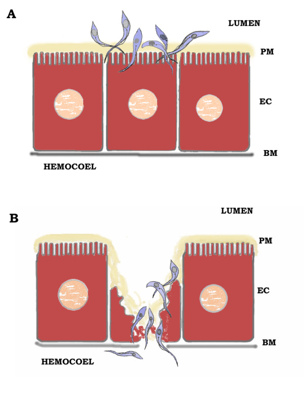
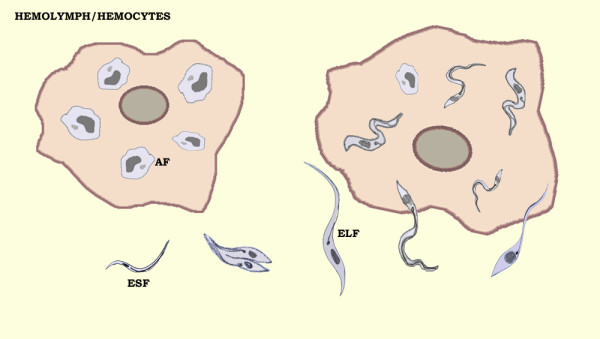
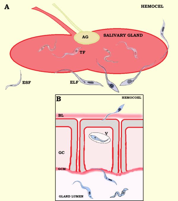
Trypanosoma rangeli is a protozoan that is non-pathogenic for humans and other mammals but causes pathology in the genus Rhodnius. T. rangeli and R. prolixus is an excellent model for studying the parasite-vector interaction, but its cycle in invertebrates remains unclear. The vector becomes infected on ingesting blood containing parasites, which subsequently develop in the gut, hemolymph and salivary glands producing short and large epimastigotes and metacyclic trypomastigotes, which are the infective forms. The importance of the T. rangeli cycle is the flagellate penetration into the gut cells and invasion of the salivary glands. The establishment of the parasite depends on the alteration of some vector defense mechanisms. Herein, we present our understanding of T. rangeli infection on the vector physiology, including gut and salivary gland invasions, hemolymph reactions and behavior alteration.


Figures

Figure 1
Figure 1
Illustration ofTrypanosoma rangeliadhesion (A) and invasion (B) toRhodnius prolixusgut epithelial cells.T. rangeli epimastigote forms bind to the epithelium cells (EC) through recognition of lectins from the extracellular membrane layers (perimicrovillar membranes – PM), for subsequent attachment and invasion. BM – basal membrane.
Figure 2
Figure 2
Illustration ofTrypanosoma rangeliinside hemocytes and in the hemolymph ofRhodnius prolixushemocoel. The parasite survives and multiplies freely in the hemolymph or penetrates into the hemocytes, especially plasmatocytes. T. rangeli epimastigote short forms (ESF) transform into amastigote forms (AF) or epimastigote long forms (ELF) which are able to invade the salivary gland.
Figure 3
Figure 3
Illustration ofTrypanosoma rangeliadhesion (A) and invasion (B) into theRhodnius prolixussalivary gland. To cross the basal lamina (BL) that surrounds the salivary glands the parasite penetrates via the flagellum and invaginates in a vacuole (V) in which the trypanosome crosses the gland cells (GC) to reach the central lumen. After reaching the gland lumen, the parasite epimastigote long forms (ELF) remain adhered to the gland cell microvilli (GCM) by their flagella, while metacyclic trypomastigote forms (TF) are found swimming freely in the saliva. AG – accessory gland; ESF – epimastigote short forms.

References

    1. Azambuja P, Garcia ES. Trypanosoma rangeliinteractions within the vectorRhodnius prolixus– a mini review. Mem Inst Oswaldo Cruz. 2005;100:567–572. - PubMed
    1. Hoare CC. Trypanosomes of Mammals, A Zoological Monograph. Blackwell, Oxford, Edinburgh; 1972.
    1. Guhl F, Vallejo GA. Trypanosoma(Herpetosoma)rangeliTejera 1920: an updated review. Mem Inst Oswaldo Cruz. 2003;98:435–442. doi: 10.1590/S0074-02762003000400001. - DOI - PubMed
    1. D ́Alessandro A. In: Biology of Kinetoplastida, vol. 1. Lamsden VHE, Evans DA, editor. Academic, London; 1976. Biology ofTrypanosoma(Herpetosoma)rangeliTejera; pp. 327–493.
    1. Marinkelle CJ. Triatoma dimidiata capitata, a natural vector ofTrypanosoma rangeliin Colombia. Rev Biol Trop. 1968;15:203–205.

Publication types

LinkOut - more resources

Cite

AltStyle によって変換されたページ (->オリジナル) /