This site needs JavaScript to work properly. Please enable it to take advantage of the complete set of features!
Skip to main page content
U.S. flag

An official website of the United States government

Dot gov

The .gov means it’s official.
Federal government websites often end in .gov or .mil. Before sharing sensitive information, make sure you’re on a federal government site.

Https

The site is secure.
The https:// ensures that you are connecting to the official website and that any information you provide is encrypted and transmitted securely.

NIH NLM Logo
Log in
Access keys NCBI Homepage MyNCBI Homepage Main Content Main Navigation
. 2012;6(5):e1644.
doi: 10.1371/journal.pntd.0001644. Epub 2012 May 8.

Inducible nitric oxide synthase in heart tissue and nitric oxide in serum of Trypanosoma cruzi-infected rhesus monkeys: association with heart injury

Affiliations

Inducible nitric oxide synthase in heart tissue and nitric oxide in serum of Trypanosoma cruzi-infected rhesus monkeys: association with heart injury

Cristiano Marcelo Espinola Carvalho et al. PLoS Negl Trop Dis. 2012.

Abstract

Background: The factors contributing to chronic Chagas' heart disease remain unknown. High nitric oxide (NO) levels have been shown to be associated with cardiomyopathy severity in patients. Further, NO produced via inducible nitric oxide synthase (iNOS/NOS2) is proposed to play a role in Trypanosoma cruzi control. However, the participation of iNOS/NOS2 and NO in T. cruzi control and heart injury has been questioned. Here, using chronically infected rhesus monkeys and iNOS/NOS2-deficient (Nos2(-/-)) mice we explored the participation of iNOS/NOS2-derived NO in heart injury in T. cruzi infection.

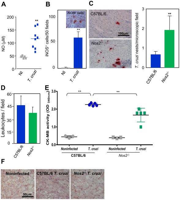
Methodology: Rhesus monkeys and C57BL/6 and Nos2(-/-) mice were infected with the Colombian T. cruzi strain. Parasite DNA was detected by polymerase chain reaction, T. cruzi antigens and iNOS/NOS2(+) cells were immunohistochemically detected in heart sections and NO levels in serum were determined by Griess reagent. Heart injury was assessed by electrocardiogram (ECG), echocardiogram (ECHO), creatine kinase heart isoenzyme (CK-MB) activity levels in serum and connexin 43 (Cx43) expression in the cardiac tissue.

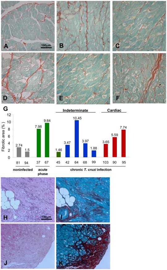
Results: Chronically infected monkeys presented conduction abnormalities, cardiac inflammation and fibrosis, which resembled the spectrum of human chronic chagasic cardiomyopathy (CCC). Importantly, chronic myocarditis was associated with parasite persistence. Moreover, Cx43 loss and increased CK-MB activity levels were primarily correlated with iNOS/NOS2(+) cells infiltrating the cardiac tissue and NO levels in serum. Studies in Nos2(-/-) mice reinforced that the iNOS/NOS2-NO pathway plays a pivotal role in T. cruzi-elicited cardiomyocyte injury and in conduction abnormalities that were associated with Cx43 loss in the cardiac tissue.

Conclusion: T. cruzi-infected rhesus monkeys reproduce features of CCC. Moreover, our data support that in T. cruzi infection persistent parasite-triggered iNOS/NOS2 in the cardiac tissue and NO overproduction might contribute to CCC severity, mainly disturbing of the molecular pathway involved in electrical synchrony. These findings open a new avenue for therapeutic tools in Chagas' heart disease.


Conflict of interest statement

The authors have declared that no competing interests exist.

Figures

Figure 1
Figure 1. Increased collagen deposits in the myocardium of T. cruzi-infected rhesus monkeys.
Collagen deposition was used to assess fibrosis in the myocardium of the left ventricle of noninfected and T. cruzi-infected monkeys. (A) Noninfected monkey #94, normal slight collagen deposits. (B) Monkey #67 (41 dpi), increased collagen deposition. (C) Monkey #45 (3 ypi), normal collagen deposits. (D) Monkey #64 (23 ypi), increased collagen deposition. (E) Monkey #99 (20 ypi) normal slight collagen deposits. (F) Monkey #95 (20 ypi), increased interstitial matrix deposits. G. Percentage of cardiac section area occupied by collagen deposits in the myocardium of the left ventricle of noninfected and T. cruzi-infected monkeys. (H–K) Serial heart sections of monkey #95 (20 ypi) showing: (H) intense infiltrates of mononuclear inflammatory cells (I) paralleling fibrosis, and (J) substitution of cardiomyocytes by mesenchymal cells in (K) an area of intense fibrosis. A–F, I and K, Picro-Sirius red stain. H and J, H&E. Bar = 100 μm.
Figure 2
Figure 2. Persistence of T. cruzi in chronically infected rhesus monkeys.
The persistence of T. cruzi parasite and antigens was evaluated by immunohistochemistry, PCR and antibody response. (A) Photomicrographs of section of myocardium of left ventricle of monkey #95 (20 ypi). Immunohistochemistry for T. cruzi antigens (black arrows and insert) associated (dotted square) or not associated (black arrow) with focal inflammation. Inflammatory infiltrates lacking parasite antigens (white arrow heads). B–C. PCR for T. cruzi kDNA (∼330 bp) in blood of noninfected controls (NI) and T. cruzi-infected rhesus monkeys at (B) 16–20 ypi and (C) 20–23 ypi. (D) PCR for T. cruzi kDNA (∼330 bp) in fragments of the left ventricle (LV) of the heart of noninfected controls and T. cruzi-infected rhesus monkeys at 20–23 ypi. Negative (−) and positive (+) controls were heart fragments of noninfected and T. cruzi-infected C57BL/6 mice, respectively. (E) Real time qPCR for the T. cruzi satellite DNA sequences Cruzi1/Cruzi2 in heart and spleen of noninfected controls and T. cruzi-infected rhesus monkeys at 20–23 ypi. (F) Standard curve of 10-fold serial dilution of DNA of epimastigote forms of the Colombian T. cruzi strain (106 to 10 parasites/mL) used for the absolute quantification by real time qPCR. The linear regression curve, coefficient of determination (r2 = 0.995) and qPCR efficiency (E = 80%) are indicated. The melting curve is also shown. (G) Serology for IgG anti-T. cruzi in rhesus monkeys prior to infection, during the acute phase (AP), when parasitemia was positive (+) and negative (−), and during the chronic phase (CP; at the end-point 20 ypi). Bar = 100 μm; Bar = 25 μm in insert in (A).
Figure 3
Figure 3. iNOS/NOS2+ cells in the myocardium and NO in the serum of T. cruzi-infected rhesus monkeys.
The presence of iNOS/NOS2+ cells in the myocardium of the left ventricles was immunohistochemically detected and NO concentration was evaluated by a Griess-based method. Photomicrograph of myocardium section of the left ventricle (A) and (B) of the noninfected monkey #81, which showed a few iNOS/NOS2+ cells. Photomicrograph of myocardioum section of the left ventricle (C) and (D) of the T. cruzi-infected monkey #95 (20 ypi). (E) Number of iNOS/NOS2+ cells in 100 microscopic fields in heart sections. (F) Concentration of NO in the serum of noninfected controls and chronically (20–23 ypi) T. cruzi-infected monkeys. (G) Concentration of NO in the serum of monkey #95 prior to infection (day 0), during the acute phase when parasitemia was positive (56 dpi) and negative (163 dpi) and during the chronic phase (16 ypi and 20 ypi). Bar = 100 μm in (A) and (C); Bar = 25 μm in (B) and (D).
Figure 4
Figure 4. Cardiomyocyte injury in T. cruzi-infected rhesus monkeys.
Cardiomyocyte damage was assessed by immunohistochemical detection of Cx43 in the myocardium of the left ventricle and CK-MB activity levels in the serum of noninfected and chronically T. cruzi-infected rhesus monkeys. (A) Photomicrograph of myocardium section of the left ventricle of the noninfected monkey #94 showing normal pattern of Cx43 expression in intercalated discs. (B) Photomicrograph of myocardium section of the left ventricle of the T. cruzi-infected monkey #64 (23 ypi) showing normal aspect. (C) Photomicrograph of left ventricle section of the T. cruzi-infected monkey #99 (20 ypi) showing normal Cx43 pattern. (D) Photomicrograph of section of left ventricle of the T. cruzi-infected monkey #90 (20 ypi) revealing Cx43 loss in myocardial area lacking inflammation. (E–F). Photomicrographs of left ventricle section of the cardiopatic T. cruzi-infected monkey #95 (20 ypi) showing Cx43 loss in area with (E) intense diffuse inflammation and (F) the substitution of cardiomyocytes by mesenchymal cells. (G) Frequency of stained Cx43 area in heart sections of noninfected and chronically T. cruzi-infected monkeys (20–23 ypi). (H) Detection of CK-MB activity in the serum of noninfected and chronically T. cruzi-infected monkeys (20–23 ypi). (I) Correlation between the number of iNOS/NOS2+ cells in heart tissue and CK-MB activity levels in serum of rhesus monkeys. Bar = 100 μm.
Figure 5
Figure 5. iNOS/NOS2 and NO status influence heart parasitism and cardiomyocyte integrity in T. cruzi-infected mice.
The mice were infected with 100 blood trypomastigotes of the Colombian T. cruzi strain and analyzed at 40 dpi. The presence of iNOS/NOS2+ cells, parasite nests, inflammatory cells and Cx43 in the myocardium was immunohistochemically detected, NO concentration was evaluated by a Griess-based method and CK-MB activity levels in the serum was biochemically determined. (A) Increased NO levels in serum of T. cruzi-infected C57BL/6 mice in comparison with noninfected controls (NI). (B) Photomicrograph of iNOS/NOS2+ cells in the cardiac tissue of infected C57BL/6 mice and quantification of iNOS/NOS2+ cells in the cardiac tissue of infected C57BL/6 mice in comparison with noninfected controls. (C) Photomicrographs and quantification of parasite nests showing increased heart parasitism in Nos2 −/− compared with T. cruzi-infected C57BL/6 mice. (D) Similar number of inflammatory cells in the heart tissue of C57BL/6 and Nos2 −/− T. cruzi-infected mice. (E) CK-MB activity levels in the serum of noninfected and T. cruzi-infected mice revealing increased CK-MB activity in T. cruzi-infected mice when compared with noninfected controls. Decreased CK-MB activity in the serum of Nos2 −/− compared with C57BL/6 T. cruzi-infected mice. (F) Preserved expression of Cx43 in the heart tissue of Nos2 −/− compared with C57BL/6 infected mice. Analysis at 40 dpi of 3–5 noninfected and 5–8 infected mice/group. * p<0.05 and ** p<0.01. Bar = 100 μm (C). Bar = 50 μm (F).

References

    1. Lannes-Vieira J, de Araújo-Jorge TC, Soeiro Mde N, Gadelha P, Corrêa-Oliveira R. The centennial of the discovery of Chagas disease: facing the current challenges. PLoS Negl Trop Dis. 2010;4:e645. - PMC - PubMed
    1. Rassi A, Jr, Rassi A, Marin-Neto JA. Chagas disease. Lancet. 2010;375:1388–1402. - PubMed
    1. Kierszenbaum F. Where do we stand on the autoimmunity hypothesis of Chagas disease? Trends Parasitol. 2005;21:513–516. - PubMed
    1. Higuchi ML, Gutierrez PS, Aiello VD, Palomino S, Bocchi E, et al. Immunohistochemical characterization of infiltrating cells in human chronic chagasic myocarditis: comparison with myocardial rejection process. Virchows Arch A Pathol Anat Histopathol. 1993;423:157–160. - PubMed
    1. Tarleton RL, Zhang L, Downs MO. "Autoimmune rejection" of neonatal heart transplants in experimental Chagas disease is a parasite-specific response to infected host tissue. Proc Natl Acad Sci USA. 1997;94:3932–3937. - PMC - PubMed

Publication types

MeSH terms

Cite

AltStyle によって変換されたページ (->オリジナル) /