This site needs JavaScript to work properly. Please enable it to take advantage of the complete set of features!
Skip to main page content
U.S. flag

An official website of the United States government

Dot gov

The .gov means it’s official.
Federal government websites often end in .gov or .mil. Before sharing sensitive information, make sure you’re on a federal government site.

Https

The site is secure.
The https:// ensures that you are connecting to the official website and that any information you provide is encrypted and transmitted securely.

NIH NLM Logo
Log in
Access keys NCBI Homepage MyNCBI Homepage Main Content Main Navigation
. 2012 May;86(10):5541-53.
doi: 10.1128/JVI.00114-12. Epub 2012 Feb 29.

Valosin-containing protein (VCP/p97) is required for poliovirus replication and is involved in cellular protein secretion pathway in poliovirus infection

Affiliations

Valosin-containing protein (VCP/p97) is required for poliovirus replication and is involved in cellular protein secretion pathway in poliovirus infection

Minetaro Arita et al. J Virol. 2012 May.

Abstract

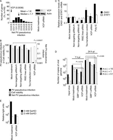
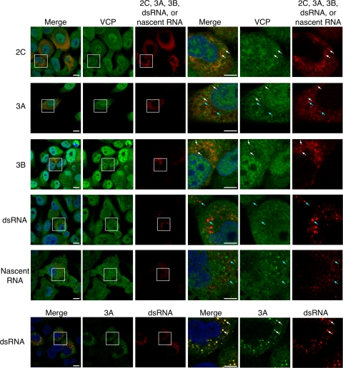
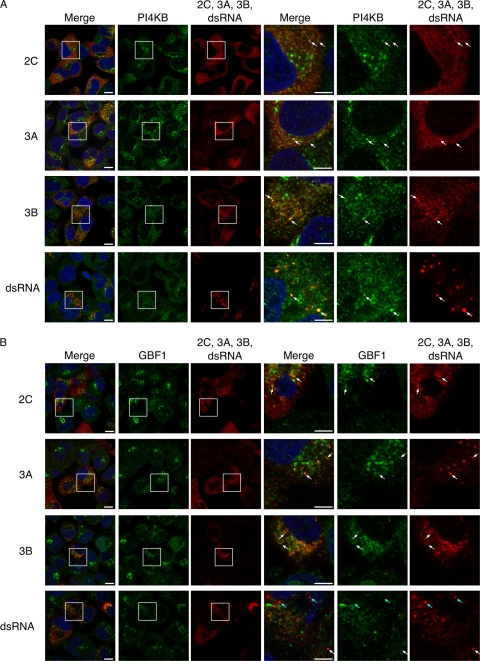
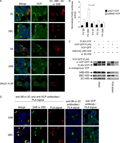
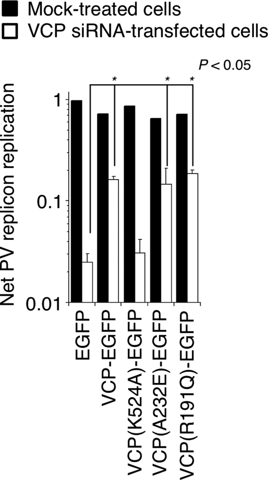
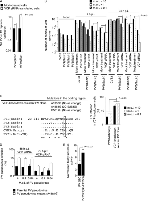
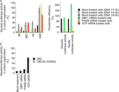
Poliovirus (PV) modifies membrane-trafficking machinery in host cells for its viral RNA replication. To date, ARF1, ACBD3, BIG1/BIG2, GBF1, RTN3, and PI4KB have been identified as host factors of enterovirus (EV), including PV, involved in membrane traffic. In this study, we performed small interfering RNA (siRNA) screening targeting membrane-trafficking genes for host factors required for PV replication. We identified valosin-containing protein (VCP/p97) as a host factor of PV replication required after viral protein synthesis, and its ATPase activity was essential for PV replication. VCP colocalized with viral proteins 2BC/2C and 3AB/3B in PV-infected cells and showed an interaction with 2BC and 3AB but not with 2C and 3A. Knockdown of VCP did not suppress the replication of coxsackievirus B3 or Aichi virus. A VCP-knockdown-resistant PV mutant had an A4881G (a mutation of E253G in 2C) mutation, which is known as a determinant of a secretion inhibition-negative phenotype. However, knockdown of VCP did not affect the inhibition of cellular protein secretion caused by overexpression of each individual viral protein. These results suggested that VCP is a host factor required for viral RNA replication of PV among membrane-trafficking proteins and provides a novel link between cellular protein secretion and viral RNA replication.


Figures

Fig 1
Fig 1
Screening of an siRNA library targeting membrane-trafficking genes for host factors of PV infection. (A) Screening of an siRNA library targeting membrane-trafficking genes. The left panel shows distribution of net PV pseudovirus infection values of siRNAs targeting 140 genes. The distribution of the numbers of genes in each range of net PV infection values is shown. Cells transfected with VCP-siRNAs showed the highest inhibitory effect on PV pseudovirus infection (0.0036 of net PV pseudovirus infection). The right panel shows Western blot analysis of VCP expression in cells treated with VCP-siRNA. (B) Inhibitory effect of VCP-siRNA treatment on PV pseudovirus infection. PV pseudovirus infection, cell viability, and net PV pseudovirus infection in siRNA-transfected cells are shown. (C) Interferon response to siRNA treatment. Expression levels of mRNAs of OAS1 and STAT1 in siRNA-transfected cells are shown. (D) Inhibitory effect of siRNA treatment targeting VCP and GBF1 on PV1 (Mahoney) infection. Cells were infected with PV1 (Mahoney) at an MOI of 10, 1, or 0.1, and then the amounts of viral genome in the cells were analyzed at 7 or 24 h p.i. Numbers of copies of viral genome of input virus and in infected cells are shown. (E) Viral protein synthesis in VCP-knockdown cells. Mock- and VCP-siRNA-treated cells (1.0 ×ばつ 104 cells) were infected with PV pseudovirus (6.5 ×ばつ 105 IU) in the presence (2 mM) or absence of GuHCl. Luciferase activities in the cells at 2 h p.i. are shown.
Fig 2
Fig 2
Localization of viral proteins, viral RNAs, and VCP in PV-infected RD cells. The first five rows show indirect immunofluorescence of viral proteins 2C, 3A, 3B, dsRNA, and VCP (nascent RNA) in PV-infected cells at 4 h p.i. The three right-most columns show magnified views of the boxed areas in the first three columns. Blue, nucleus (staining with Hoechst 33342); green, VCP; red, 2C, 3A, 3B, dsRNA, or nascent viral RNA. Scale bars, 10 μm (first three columns) and 5 μm (three right-most columns). The bottom row shows indirect immunofluorescence images of viral proteins 3A and dsRNA in PV-infected cells. The columns are as described above. Blue, nucleus (staining with Hoechst 33342); green, 3A; red, dsRNA. Scale bars, 10 μm (first three panels) and 5 μm (three right-most panels). White arrows indicate some of the colocalized sites, and cyan arrows indicate some of the noncolocalized sites.
Fig 3
Fig 3
Localization of viral proteins, viral RNAs, and PI4KB and GBF1 in PV-infected RD cells. Indirect immunofluorescence images of PI4KB (A) or GBF1 (B) are shown. The three right-most columns are magnified views of the boxed areas in the first three columns. Blue, nucleus (staining with Hoechst 33342); green, PI4KB or GBF1; red, 2C, 3A, 3B, or dsRNA. Scale bars, 10 μm (first three columns) and 5 μm (three right-most columns). White arrows indicate some of the colocalized sites, and cyan arrows indicated some of the noncolocalized sites.
Fig 4
Fig 4
Localization of overexpressed viral proteins with VCP. (A) Localization of VCP in HEK293 cells expressing 2C, 2BC, 3A, and 3AB mutants. Blue, nucleus (staining with Hoechst 33342); green, VCP; red, 2C, 2BC, 3A, 3AB, or a 3AB mutant. Scale bar, 10 μm. (B) Mammalian two-hybrid assay for VCP and PV proteins. Closed bars, pACT-VCP and pBIND-PV proteins; open bars, pBIND-VCP and pACT-PV proteins. Normalized firefly luciferase activities are shown. (C) Immunoprecipitation of 3AB, 2BC, and 2C with VCP. HEK293 cells expressing 3AB, 2BC, or 2C (3AB-His, 2BC-His, or 2C-His) with FLAG-VCP, VCP-GFP-FLAG, or VCP-GFP were precipitated by anti-FLAG antibody. The amounts of VCP and viral proteins in input (4% of cell lysate used for immunoprecipitation) and pulldowns (immunoprecipitated with anti-FLAG antibody) were analyzed by Western blotting with anti-VCP antibody (for FLAG-VCP, VCP-GFP-FLAG, and VCP-GFP) or anti-Penta-His antibody (for 3AB-His, 2BC-His, and 2C-His). (D) PLA signals between viral proteins and VCP in 3AB- and 2BC-expressing cells. 3AB- and 2BC-expressing cells were incubated with anti-3B/2C antibodies with anti-VCP antibody (left six panels) or with individual antibodies (anti-3B/2C antibodies or anti-VCP antibody) and then subjected to PLA. Blue, nucleus (staining with 4′,6′-diamidino-2-phenylindole[DAPI]); green; 3AB or 2BC or VCP; red, PLA signals.
Fig 5
Fig 5
Role of ATPase activity of VCP in PV replication. PV replicon replication was analyzed in mock-treated and VCP-siRNA-transfected HEK293 cells expressing EGFP (control) or VCP mutants. A t test was performed for the net PV replicon replication between EGFP-expressing cells (control) and cells expressing each of the VCP mutants.
Fig 6
Fig 6
VCP act as a PV-specific host factor of viral replication via a cellular secretion pathway. (A) Effect of VCP-siRNA treatment on PV and AV replicon replication. Luciferase activity in mock-treated and VCP-siRNA-transfected cells was analyzed at 7 h p.t. of RNA transcripts of each replicon. (B) Effect of VCP-siRNA treatment on CVB3 and type 1, 2, and 3 PV infection. Cells were infected with CVB3 or type 1, 2, and 3 PV (Sabin) at an MOI of 10, 1, or 0.1, and then the amounts of viral genome in the infected cells were analyzed at 7 or 24 h p.i. Numbers of copies of the viral genome of input virus and in infected cells are shown. The number of copies of the viral genome in VCP-siRNA-treated cells was compared with that in mock-treated cells infected with viruses at each MOI. (C) Isolation of PV mutant resistant to VCP knockdown. The left panel shows mutations in the coding region of a VCP-knockdown-resistant PV clone (top) and partial 2C sequences around aa 253 (aa 241 to 257 of 2C) of enteroviruses (bottom) are shown. Amino acid 253 is highlighted in a rectangle. Results of a PV infection assay in VCP-knockdown cells are shown at right. VCP-siRNA-transfected or mock-treated HEK293 cells were infected with PV1 (Mahoney) or a VCP-knockdown-resistant PV clone at 48 h p.t. of VCP siRNA at an MOI of 10, 1, and 0.1. Numbers of copies of viral RNA were determined at 7 h p.i. PV infection in VCP-knockdown cells is presented as a percentage of the number of copies of viral RNA in VCP-siRNA-transfected cells compared to that in mock-treated cells (100%). A t test was performed between samples for each MOI. (D) Effect of VCP-siRNA treatment on PV pseudovirus infection with an A4881G mutation. VCP-siRNA-transfected HEK293 cells were infected with a parental PV pseudovirus or a PV pseudovirus mutant with an A4881G mutation at an MOI of 4, 0.4, or 0.04 at 48 or 72 h p.t. of VCP-siRNA. Percent PV pseudovirus infection is shown. (E) Mammalian two-hybrid assay for VCP (pBIND-VCP) and PV proteins [pACT-PV 2BC or PV 2BC(2C-E253G)]. Normalized firefly luciferase activities are shown.
Fig 7
Fig 7
Effect of knockdown of VCP on the inhibition of cellular protein secretion caused by overexpression of each individual viral protein. (A) Inhibition of cellular protein secretion caused by the individual viral protein in siRNA-treated cells. Secretion of Gaussia luciferase was analyzed in mock-treated and GBF1-, PI4KB-, and VCP-siRNA-transfected HEK293 cells expressing viral proteins 2B, 2BC, 3A, and 3AB (left). For mock-treated cells, DNAs (control vector and Gaussia luciferase expression vector) diluted by 1-, 4-, and 8-fold were transfected to control the transfection efficiency. Gaussia luciferase activity in cells transfected with a control expression vector was taken as 100%. At right is an evaluation of DNA transfection efficiency. Gaussia luciferase activity in the cells transfected with a control vector and Gaussia luciferase expression vector and Renilla luciferase activity in the cells transfected with Renilla luciferase expression vector are shown as percentages. (B) Effect of a 2C-E253G mutation on the inhibition of cellular protein secretion caused by overexpression of 2BC protein in siRNA-treated cells. Gaussia luciferase activity was determined in siRNA-transfected cells, with the value for cells transfected with a control expression vector taken as 100%.

References

    1. Arita M, Ami Y, Wakita T, Shimizu H. 2008. Cooperative effect of the attenuation determinants derived from poliovirus Sabin 1 strain is essential for attenuation of enterovirus 71 in the NOD/SCID mouse infection model. J. Virol. 82:1787–1797 - PMC - PubMed
    1. Arita M, Horie H, Arita M, Nomoto A. 1999. Interaction of poliovirus with its receptor affords a high level of infectivity to the virion in poliovirus infections mediated by the Fc receptor. J. Virol. 73:1066–1074 - PMC - PubMed
    1. Arita M, et al. 2011. Phosphatidylinositol 4-kinase III beta is a target of enviroxime-like compounds for antipoliovirus activity. J. Virol. 85:2364–2372 - PMC - PubMed
    1. Arita M, Nagata N, Sata T, Miyamura T, Shimizu H. 2006. Quantitative analysis of poliomyelitis-like paralysis in mice induced by a poliovirus replicon. J. Gen. Virol. 87:3317–3327 - PubMed
    1. Arita M, Takebe Y, Wakita T, Shimizu H. 2010. A bifunctional anti-enterovirus compound that inhibits replication and early stage of enterovirus 71 infection. J. Gen. Virol. 91:2734–2744 - PubMed

Publication types

LinkOut - more resources

Cite

AltStyle によって変換されたページ (->オリジナル) /