This site needs JavaScript to work properly. Please enable it to take advantage of the complete set of features!
Skip to main page content
U.S. flag

An official website of the United States government

Dot gov

The .gov means it’s official.
Federal government websites often end in .gov or .mil. Before sharing sensitive information, make sure you’re on a federal government site.

Https

The site is secure.
The https:// ensures that you are connecting to the official website and that any information you provide is encrypted and transmitted securely.

NIH NLM Logo
Log in
Access keys NCBI Homepage MyNCBI Homepage Main Content Main Navigation
. 2012 Mar 15;205(6):1019-23.
doi: 10.1093/infdis/jir874. Epub 2012 Jan 25.

Infection with Cryptosporidium hominis provides incomplete protection of the host against Cryptosporidium parvum

Affiliations

Infection with Cryptosporidium hominis provides incomplete protection of the host against Cryptosporidium parvum

Abhineet Sheoran et al. J Infect Dis. .

Abstract

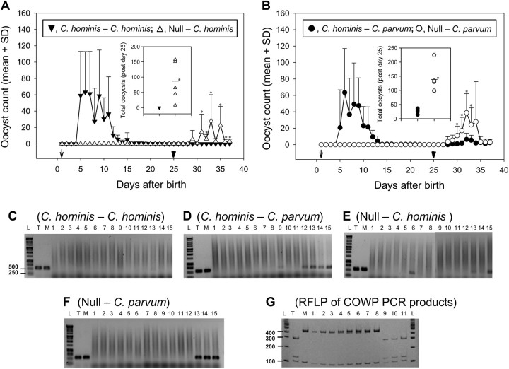
Cryptosporidium hominis and Cryptosporidium parvum, which infect humans equally, are genetically/antigenically almost identical. It remains unclear, however, whether infection with C. hominis protects against C. parvum. Gnotobiotic piglets were used to investigate cross-protection. After ≥3 days of recovery from C. hominis infection, the piglets were completely protected against subsequent challenge with C. hominis but only partially against challenge with C. parvum, as compared with age-matched control animals challenged with either species. In conclusion, C. hominis-specific immunity was sufficient to completely protect against challenge with the same species but insufficient to provide the same level of protection against C. parvum.


Figures

Figure 1.
Figure 1.
Cross-protection studies in the gnotobiotic piglet model. A, B, Piglets shed oocysts in feces following primary infection (arrows) with Cryptosporidium hominis (C. hominis–C. hominis, A; and C. hominis–Cryptosporidium parvum, B). Piglets that were not subjected to primary infection remained negative until infected (null–C. hominis, A; and null–C. parvum, B). Following challenge infection (arrowheads), piglets of all groups (A and B) except the C. hominis–C. hominis group (A) shed oocysts. The y-axis shows mean + standard deviation (SD) of oocyst count (in 30 microscopic fields at ×ばつ100 magnification). The statistically significant differences of oocyst count between the groups on a particular day are shown with asterisks. Insets of figure A and B show scatterplots (means represented by a bar) of total oocyst counts of each piglet during 12 days of postchallenge (days 26–37 after birth) from null–C. hominis/C. hominisC. hominis groups and null–C. parvum/C. parvumC. parvum groups, respectively. Statistically significant differences of total oocyst count between groups are indicated with asterisks. C–G, Cryptosporidium oocyst wall protein (COWP) polymerase chain recation (PCR) restriction fragment length polymorphism (RFLP) analysis of intestinal sections from a piglet of each of the 4 infection groups at necropsy 12 days after challenge infection. CF, PCR analysis of the 15 intestinal sections; lanes 1–12 correspond to sections of the small intestine and lanes 13–15 to the large intestine. G, RFLP results of the 553-bp amplicon digested with RsaI. Lanes T and M are positive controls with C. hominis TU502 and C. parvum MD DNA, respectively. Lane L contains 1-kb ladder (C–F) or 100-bp ladder (G). The experimental group for panels CF is shown above each figure. G, Lanes 1–5 contain RFLP products of the COWP PCR products of lanes 9 and 12–15 of the C. hominisC. parvum group, respectively; lanes 6–8 contain COWP PCR-RFLP products of lanes 13–15 of the null–C. parvum group, respectively; and lanes 9–11 contain RFLP products of the COWP PCR products of lanes 6, 13, and 15 of the null–C. hominis group, respectively. Digestion with RsaI yields fragments of 285, 125, 106, and 34 bp for type C. hominis and 410, 106, and 34 bp for C. parvum. The 34-bp fragment is not shown as it runs through the gel.
Figure 2.
Figure 2.
Immunoblot reactivity of serum immunoglobulin G (IgG) and mucosal immunoglobulin A (IgA) (2 animals from each group) collected at necropsy 12 days after challenge infection with antigens of Cryptosporidium hominis TU502 (lane T) and Cryptosporidium parvum MD (lane M). A 105-kDa antigen of C. hominis and a 100-kDa antigen of C. parvum are indicated by the arrow and arrowhead, respectively. Molecular weight markers are shown on the left, and molecular weights of some of the Cryptosporidium antigens are on the right.

References

    1. Tzipori S, Widmer G. The biology of Cryptosporidium. Contrib Microbiol. 2000;6:1–32. - PubMed
    1. Sturbaum GD, Schaefer DA, Jost BH, Sterling CR, Riggs MW. Antigenic differences within the Cryptosporidium hominis and Cryptosporidium parvum surface proteins P23 and GP900 defined by monoclonal antibody reactivity. Mol Biochem Parasitol. 2008;159:138–41. - PubMed
    1. Morgan-Ryan UM, Fall A, Ward LA, et al. Cryptosporidium hominis n. sp. (Apicomplexa: Cryptosporidiidae) from Homo sapiens. J Eukaryot Microbiol. 2002;49:433–40. - PubMed
    1. Xu P, Widmer G, Wang Y, et al. The genome of Cryptosporidium hominis. Nature. 2004;431:1107–12. - PubMed
    1. Akiyoshi DE, Feng X, Buckholt MA, Widmer G, Tzipori S. Genetic analysis of a Cryptosporidium parvum human genotype 1 isolate passaged through different host species. Infect Immun. 2002;70:5670–5. - PMC - PubMed

Publication types

MeSH terms

Cite

AltStyle によって変換されたページ (->オリジナル) /