This site needs JavaScript to work properly. Please enable it to take advantage of the complete set of features!
Skip to main page content
U.S. flag

An official website of the United States government

Dot gov

The .gov means it’s official.
Federal government websites often end in .gov or .mil. Before sharing sensitive information, make sure you’re on a federal government site.

Https

The site is secure.
The https:// ensures that you are connecting to the official website and that any information you provide is encrypted and transmitted securely.

NIH NLM Logo
Log in
Access keys NCBI Homepage MyNCBI Homepage Main Content Main Navigation
. 2011 Jul 10;18(8):908-14.
doi: 10.1038/nsmb.2088.

Dimerization of Plasmodium vivax DBP is induced upon receptor binding and drives recognition of DARC

Affiliations

Dimerization of Plasmodium vivax DBP is induced upon receptor binding and drives recognition of DARC

Joseph D Batchelor et al. Nat Struct Mol Biol. .

Abstract

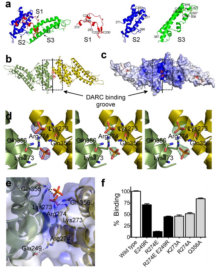
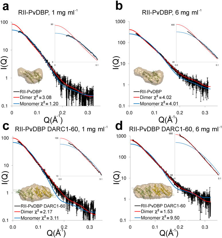
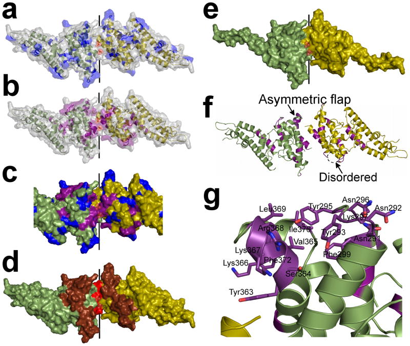
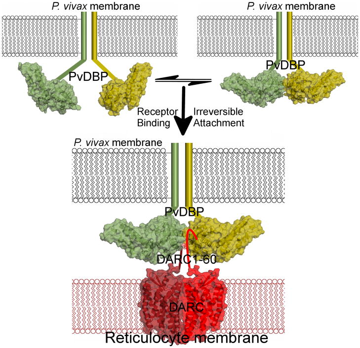
Plasmodium vivax and Plasmodium knowlesi invasion depends on the parasite Duffy-binding protein DBL domain (RII-PvDBP or RII-PkDBP) engaging the Duffy antigen receptor for chemokines (DARC) on red blood cells. Inhibition of this key interaction provides an excellent opportunity for parasite control. There are competing models for whether Plasmodium ligands engage receptors as monomers or dimers, a question whose resolution has profound implications for parasite biology and control. We report crystallographic, solution and functional studies of RII-PvDBP showing that dimerization is required for and driven by receptor engagement. This work provides a unifying framework for prior studies and accounts for the action of naturally acquired blocking antibodies and the mechanism of immune evasion. We show that dimerization is conserved in DBL-domain receptor engagement and propose that receptor-mediated ligand dimerization drives receptor affinity and specificity. Because dimerization is prevalent in signaling, our studies raise the possibility that induced dimerization may activate pathways for invasion.


Figures

Figure 1
Figure 1
RII-PvDBP is composed of three subdomains, and a sulfotyrosine pocket within a DARC binding groove is formed by the RII-PvDBP dimer. (a) RII-PvDBP separated into the three subdomains. Subdomain 1 (S1 - red) contains the β-hairpin, subdomain 2 (S2 - blue) is a four helix bundle and subdomain 3 (S3 -green) forms a second helical bundle. (b) The RII-PvDBP dimer in ribbon representation, rotated by 180° along x. Monomers are in green and yellow. The DARC binding groove is outlined by a dashed box and the dimer interface is indicated by a solid line. (c) Electrostatic mapping of the RII-PvDBP dimer, rotated by 180° along x. The DARC binding groove is positively charged. (d) Density which clearly identifies selenate or phosphate at the RII-PvDBP dimer interface is shown. Left - Selenium anomalous difference map (blue mesh) from crystals grown in the presence of selenate, contoured at 4σ. Middle - the difference map (green mesh), contoured at 2.5σ, of crystals grown in phosphate, prior to addition of phosphates. Right - the omit map (green mesh), contoured at 2.5σ, of the final refined structure with the phosphates omitted. Phosphates are drawn in stick and colored red and yellow. Side chains of residues involved in interactions are shown in stick. (e) The putative sulfotyrosine binding pocket and the Arg274-Glu249 salt bridge shown. Phosphates are drawn in stick and colored red and yellow. Side chains of residues involved in interactions are shown in stick and contacts are depicted by dashed black lines. (f) Percentage of cells expressing point mutants of RII-PvDBP that bind RBCs relative to wildtype, shown with standard error. A paired two-tailed student t-test indicated that all mutants compared to wildtype have a p-value<0.0001. White bar – wildtype. Black bars – dimer mutants and rescue. Grey bars – sulfotyrosine binding mutants.
Figure 2
Figure 2
DARC binding drives dimerization of RII-PvDBP. Experimental (black) and theoretical SAXS plots for the monomer (blue) and dimer (red) at different concentrations. An expanded plot of the low-angle data (0 < Q < 0.1) that clearly delineates oligomeric state is shown in the top right insert. Ab initio reconstructions are overlayed on structures (bottom left insert) with monomers colored in green and yellow and molecular envelopes in sand. (a) RII-PvDBP at 1 mg ml−1. (b) RII-PvDBP at 6 mg ml−1. (c) RII-PvDBP–DARC1–60 at 1 mg ml−1. (d) RII-PvDBP–DARC1–60 at 6 mg ml−1.
Figure 3
Figure 3
The sulfotyrosine pocket, DARC binding groove and dimer interface are under selective pressure and are targeted by blocking-antibodies. Monomers are in green and yellow. (a) Polymorphic residues, (blue) are excluded from the dimer interface but evenly distributed over the remaining RII-PvDBP surface. (b) Amino acid substitutions which abrogate RBC rosetting, (purple) map to the DARC binding groove and dimer interface. (c) Overlay of polymorphic residues (blue) and critical receptor binding residues (purple) on the dimer. The DARC binding groove at the dimer interface is composed of essential residues and devoid of polymorphisms. (d) Epitopes recognized by blocking-antibodies (red – most significant, brown – significant) map to the functional regions of RII-PvDBP which include the dimer interface and DARC binding groove. (e) The minimal binding domain of RII-PvDBP (residues 256–426) contains the full dimer interface and DARC binding groove. (f) A global view of the dimer which shows the asymmetric flap is disordered in chain A. Essential residues are colored in purple. (g) A detailed view of the asymmetric flap shows this region contains several essential residues suggesting a second potential DARC binding site.
Figure 4
Figure 4
RII-PvDBP’s dimer interface and receptor binding site are conserved in VAR2CSA DBL6ε. (a) Examination of the crystal packing interfaces for VAR2CSA DBL6ε revealed a dimeric organization identical to the RII-PvDBP dimer. Critical VAR2CSA DBL6ε binding residues are shown in red and map to the putative sulfotyrosine binding pocket indicating that both the receptor binding pocket and dimer interface are conserved in these two DBL domains. Monomers are colored green/yellow in both cases. Top panel – reported asymmetric unit for DBL6ε, Middle panel – reorganized dimer based solely on crystal symmetry, Bottom panel – RII-PvDBP dimer. (b) Overlay of RII-PvDBP (green) and DBL6ε (brown) reveals critical binding residues for each protein superpose well (Lys273 and Arg274 from RII-PvDBP, and Lys2392 and Lys2395 from DBL6ε).
Figure 5
Figure 5
PvDBP binds DARC via a model of receptor-mediated ligand-dimerization. PvDBP exists as an equilibrium of monomers and dimers that is shifted to dimerization upon receptor-binding. RII-PvDBP monomers are in green/yellow. The P. vivax membrane is in black and the reticulocyte membrane is in red. Flat lines represent portions of PvDBP not in the crystal structure. The DARC homodimer is represented by the crystal structure of a related GPCR, CXCR4’s, homodimeric membrane spanning region, in dark/light red. DARC1–60 is shown as a flat line. Two PvDBP molecules bind two DARC molecules as indicated by our stoichiometry measurements.

References

    1. Mason SJ, Miller LH, Shiroishi T, Dvorak JA, McGinniss MH. The Duffy Blood Group Determinants: Their Role in the Susceptibility of Human and Animal Erythrocytes to Plasmodium knowlesi Malaria. British Journal of Haematology. 1977;36:327–335. - PubMed
    1. Miller L, Mason S, Clyde D, McGinniss M. The resistance factor to Plasmodium vivax in blacks. The Duffy-blood-group genotype, FyFy. N Engl J Med. 1976;295:302–304. - PubMed
    1. Miller LH, Mason SJ, Dvorak JA. Erythrocytes receptors for (Plasmodium knowlesi) malaria: Duffy blood group determinants. Science. 1975;189:561–563. - PubMed
    1. Adams JH, et al. A family of erythrocyte binding proteins of malaria parasites. Proc Natl Acad Sci U S A. 1992;89:7085–9. - PMC - PubMed
    1. Chitnis C, Miller L. Identification of the erythrocyte binding domains of Plasmodium vivax and Plasmodium knowlesi proteins involved in erythrocyte invasion. J Exp Med. 1994;180:497–506. - PMC - PubMed

Publication types

Substances

Associated data

Cite

AltStyle によって変換されたページ (->オリジナル) /