This site needs JavaScript to work properly. Please enable it to take advantage of the complete set of features!
Skip to main page content
U.S. flag

An official website of the United States government

Dot gov

The .gov means it’s official.
Federal government websites often end in .gov or .mil. Before sharing sensitive information, make sure you’re on a federal government site.

Https

The site is secure.
The https:// ensures that you are connecting to the official website and that any information you provide is encrypted and transmitted securely.

NIH NLM Logo
Log in
Access keys NCBI Homepage MyNCBI Homepage Main Content Main Navigation
. 2011;5(5):e1141.
doi: 10.1371/journal.pntd.0001141. Epub 2011 May 31.

Effect of BMAP-28 antimicrobial peptides on Leishmania major promastigote and amastigote growth: role of leishmanolysin in parasite survival

Affiliations

Effect of BMAP-28 antimicrobial peptides on Leishmania major promastigote and amastigote growth: role of leishmanolysin in parasite survival

Miriam A Lynn et al. PLoS Negl Trop Dis. 2011.

Abstract

Background: Protozoan parasites, such as Leishmania, still pose an enormous public health problem in many countries throughout the world. Current measures are outdated and have some associated drug resistance, prompting the search into novel therapies. Several innovative approaches are under investigation, including the utilization of host defence peptides (HDPs) as emerging anti-parasitic therapies. HDPs are characterised by their small size, amphipathic nature and cationicity, which induce permeabilization of cell membranes, whilst modulating the immune response of the host. Recently, members of the cathelicidin family of HDPs have demonstrated significant antimicrobial activities against various parasites including Leishmania. The cathelicidin bovine myeloid antimicrobial peptide 28 (BMAP-28) has broad antimicrobial activities and confers protection in animal models of bacterial infection or sepsis. We tested the effectiveness of the use of BMAP-28 and two of its isomers the D-amino acid form (D-BMAP-28) and the retro-inverso form (RI-BMAP-28), as anti-leishmanial agents against the promastigote and amastigote intracellular Leishmania major lifecycle stages.

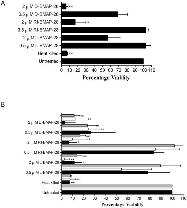
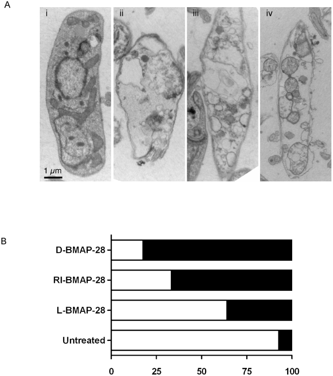
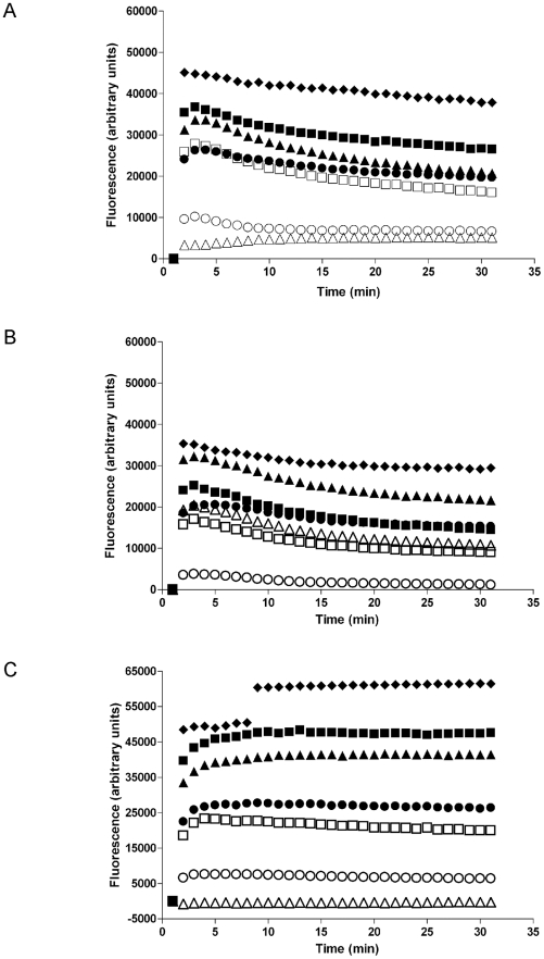
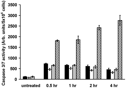
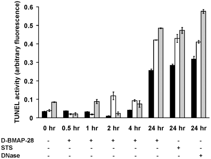
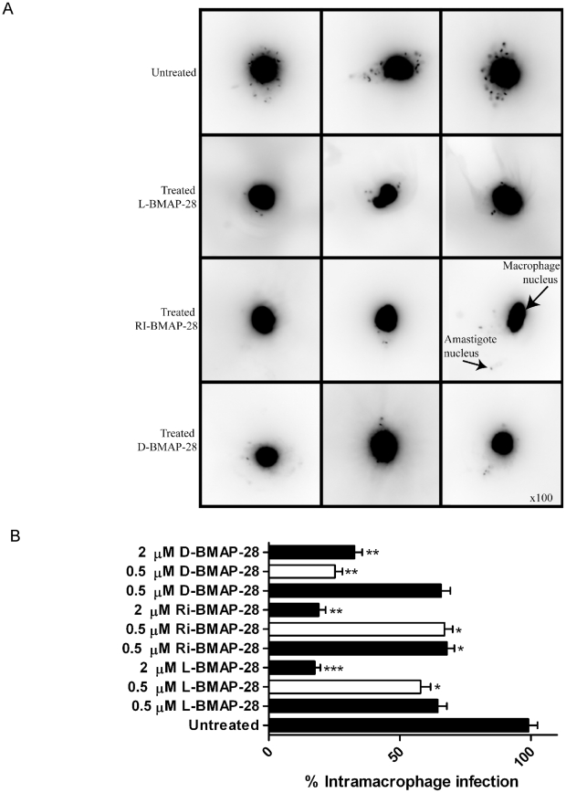
Methodology/principal findings: An MTS viability assay was utilized to show the potent antiparasitic activity of BMAP-28 and its protease resistant isomers against L. major promastigotes in vitro. Cell membrane permeability assays, caspase 3/7, Tunel assays and morphologic studies suggested that this was a late stage apoptotic cell death with early osmotic cell lysis caused by the antimicrobial peptides. Furthermore, BMAP-28 and its isomers demonstrated anti-leishmanial activities against intracellular amastigotes within a macrophage infection model.

Conclusions/significance: Interestingly, D-BMAP-28 appears to be the most potent antiparasitic of the three isomers against wild type L. major promastigotes and amastigotes. These exciting results suggest that BMAP-28 and its protease resistant isomers have significant therapeutic potential as novel anti-leishmanials.


Conflict of interest statement

The authors have declared that no competing interests exist.

Figures

Figure 1
Figure 1. MTS viability assay of Leishmania major strains when treated with BMAP-28 variants.
(A) L. major Friedlin strain was treated with 0.5 μM and 2 μM concentrations of L-, RI- and D-BMAP-28 for 4 hours. (B) L. major Seidman strain wild-type (wt, filled bars), GP63 knockout (ko, clear bars) and reconstituted (ko+, grey bars) were treated with 0.5 μM and 2 μM concentrations of L-, RI- and D-BMAP-28 for 4 hours. Cell viability was assessed at 490 nm and expressed as a percentage of untreated control cells. Three complete biological replicates were performed and the standard errors are shown. Paired T-tests untreated vs treated indicated significance, where *p<0.05, **p<0.005, ***p<0.0005. The bar graph was created using GraphPad prism 4.
Figure 2
Figure 2. Analyses of effect of BMAP-28 peptides on L. major cells.
(A) Wild-type cells were (i) untreated or treated with (ii) L-BMAP-28, (iii) RI-BMAP-28 or (iv) D-BMAP-28 at 0.5 μM concentrations for 4 hours prior to fixation for TEM analyses. The figure shows cross-sections of cells at ×ばつ20,000. Notice that the untreated cell has an intact membrane, an intact cytosol and a few small vacuoles. The treated cells however possess disrupted membranes, large fluid filled vacuoles and loss of cytosolic contents; indicative of osmotic lysis. (B) Quantitative analyses of intact (clear) versus damaged (filled) cells were carried out on wt cells in presence/absence of treatment to assess the extent of the damage. Damage was assessed based on three criterion, membrane disruption, loss of cytosolic contents and presence of large fluid filled vacuoles, 120 cells were analysed by TEM for each condition.
Figure 3
Figure 3. Leishmania cell membrane permeabilization by L-, D- and RI-BMAP-28.
L. major cell lines were treated with 0.5 μM (clear symbols) or 2.0 μM (filled symbols) of each of the three BMAP-28 isomers, L-BMAP-28 (▴, しろさんかく), RI-BMAP-28 (しろまる, •) and D-BMAP-28 (しろいしかく, ▪) and analyzed for membrane permeabilization using SYTOX. (A) L. major wt, (B) L. major ko, (C) L. major ko+. Incubation of cells with 0.5% Triton X-100 (filled diamonds) were used as a positive control. Three complete biological replicates were performed and the average is shown, with standard deviations less than 5% for all values.
Figure 4
Figure 4. Induction of early apoptosis events in L. major strains after incubation with D-BMAP-28.
L. major wt (black bars), L. major ko (clear bars) and L. major ko+ (grey bars) were treated with 2 μM D-BMAP-28 for 0.5 hr, 1 hr, 2 hr, and 4 hr and then analyzed for caspase 3/7 activation. L. major wt cells treated with 16 μM staurosporine for the indicated period of time were used as positive control (striped bars). Each bar represents the mean of four replicates; error bar represents the standard error of the mean.
Figure 5
Figure 5. Induction of late apoptosis events in L. major strains after incubation with D-BMAP-28.
Quantitative colorimetric analysis of DNA degradation in L. major wt (black bars), L. major ko (clear bars) and L. major ko+ (grey bars) after treatment with 2 μM D-BMAP-28 for 0.5 hr, 1 hr, 2 hr, 4 hr and 24 hr using the TUNEL assay. Cells treated with 16 μM staurosporine (STS) as well as DNase treated cell lysates were used as positive controls. Each bar represents the mean of three replicates; error bar represents the standard error of the mean.
Figure 6
Figure 6. The effect of BMAP-28 isomers on intramacrophage L. major infection.
(A) The figure displays images that were collected from peritoneal macrophages infected with L. major Seidman wt strains for 24 h, and treated with L-, RI- and D-BMAP-28 at 0.5 μM for an additional 24 h. Infected macrophages were stained with DAPI and examined under UV light with an upright fluorescent microscope, using 100 magnification. (B) Peritoneal macrophages were infection with L. major Seidman wt (filled) and ko strains (open) for 24 h, and treated with L-, RI- and D-BMAP-28 at 0.5 and/or 2 μM for 48 h. Infections were stained with DAPI and parasite burden was quantified as an average of 100 macrophages, and expressed as a percentage of the control infection. The average number of three complete biological replicates as well as the standard errors are shown. Paired T-tests untreated vs treated indicated significance, where * p<0.05, **p<0.005, ***p<0.0005.

Comment in

References

    1. McConville MJ, Handman E. The molecular basis of Leishmania pathogenesis. Int J Parasitol. 2007;37:1047–1051. - PubMed
    1. Marsden PD. Mucosal leishmaniasis ("espundia" Escomel, 1911). Trans R Soc Trop Med Hyg. 1986;80:859–876. - PubMed
    1. Sacks D, Anderson C. Re-examination of the immunosuppressive mechanisms mediating non-cure of Leishmania infection in mice. Immunol Rev. 2004;201:225–238. - PubMed
    1. Besteiro S, Williams RA, Coombs GH, Mottram JC. Protein turnover and differentiation in Leishmania. Int J Parasitol. 2007;37:1063–1075. - PMC - PubMed
    1. Chakravarty J, Sundar S. Drug Resistance in Leishmaniasis. J Glob Infect Dis. 2010;2:167–176. - PMC - PubMed

Publication types

Cite

AltStyle によって変換されたページ (->オリジナル) /