Review
doi: 10.1038/gt.2011.50.
Epub 2011 Apr 28.
MicroRNA therapeutics
Affiliations
- PMID: 21525952
- PMCID: PMC3237828
- DOI: 10.1038/gt.2011.50
Item in Clipboard
Review
MicroRNA therapeutics
J A Broderick et al.
Gene Ther.
2011 Dec.
Display options
Format
Display options
Format
Abstract
MicroRNAs (miRNAs) provide new therapeutic targets for many diseases, while their myriad roles in development and cellular processes make them fascinating to study. We still do not fully understand the molecular mechanisms by which miRNAs regulate gene expression nor do we know the complete repertoire of mRNAs each miRNA regulates. However, recent progress in the development of effective strategies to block miRNAs suggests that anti-miRNA drugs may soon be used in the clinic.
Figures
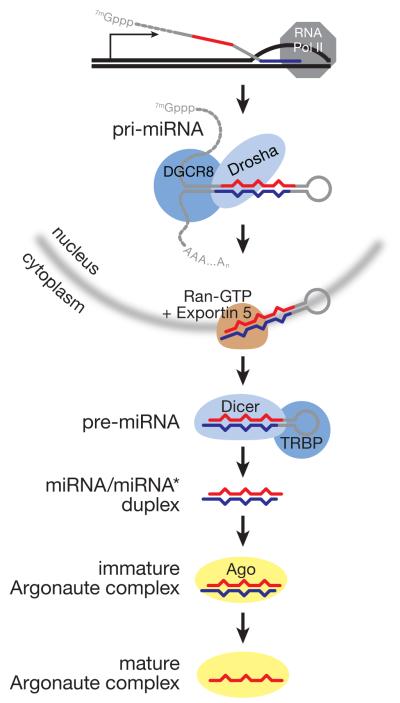
miRNA biogenesis in mammals.
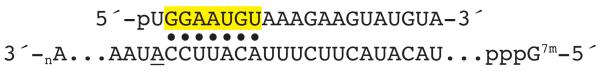
miRNAs bind target mRNAs via their seed sequence. Typical miRNA-binding sites also feature an adenosine (underlined) across from the first nucleotide of the miRNA, even though the structure of a miRNA bound to an Argonaute protein precludes base pairing at this position.
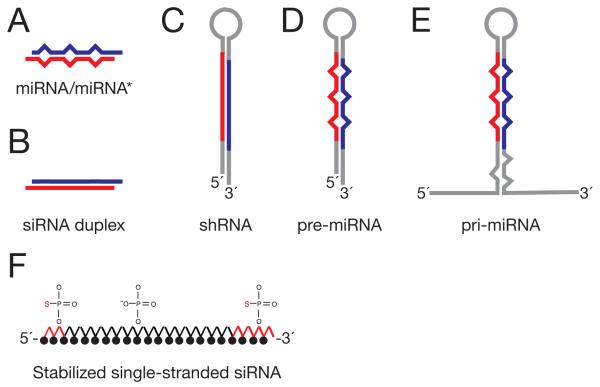
miRNA replacement strategies: (A) mature miRNA/miRNA* duplex; (B) small interfering RNA duplex; (C) small hairpin RNA; (D) pre-miRNA; (E) pri-miRNA; (F) modified single stranded RNA.
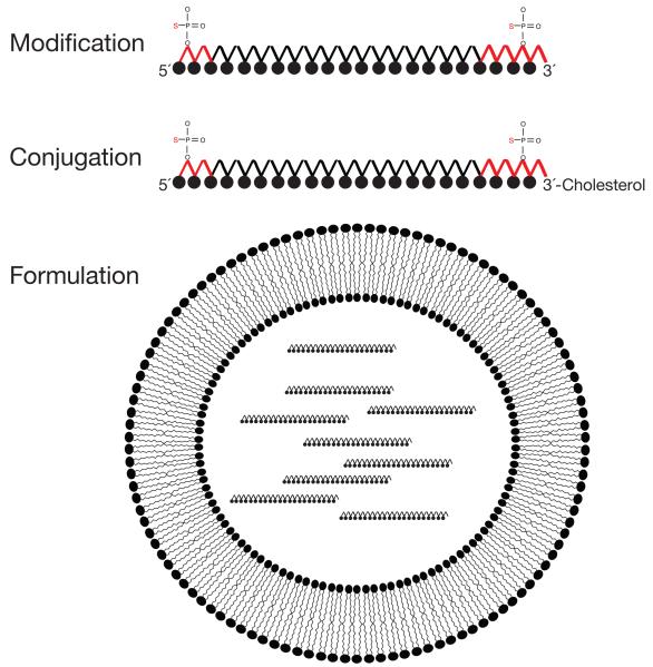
Strategies for delivery of anti-miRNA oligonucleotides to cells in vivo. (A) Modification. Black filled circles represent 2′-O-methyl, 2′-O-methoxyethyl, or 2′-fluoro modified nucleotides. (B) Conjugation. Antagomirs are 2′-O-methyl oligonucleotides conjugated to cholesterol at their 3′ ends, and contain phosphorothioate linkages between nucleotides at both ends in place of natural phosphate linkages. (C) Formulation. Lipid nanoparticles are lipid vesicles containing therapeutic RNA. The formulated lipid bilayer encapsulates the therapeutic RNA, delivering it to cells and promoting fusion with the phospholipid bilayer of cell membranes. Individual lipids within the vesicle bilayer can contain ionizable head groups that will disrupt the endosome at low pH to release the therapeutic RNA to the cytoplasm.
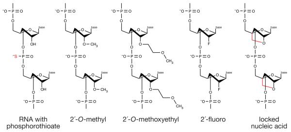
Chemical modifications that improve the stability, biodistribution, and delivery of ASOs. RNA (red; S indicates sulfur substitution of a non bridging oxygen to make a phosphorothioate linkage between nucleotides), 2′-O-methyl RNA contains a methyl group bound to the 2′ oxygen of the ribose; 2′-O-methoxyethyl RNA contains a methoxy group bound to the 2′ oxygen of the ribose; 2′-fluoro RNA contains fluorine molecule bound to the 2′ oxygen of the ribose; and locked nucleic acid (red) introduces a 2′,4′ methylene bridge in the ribose to form a bicyclic nucleotide).
References
-
- Medina PP, Slack FJ. microRNAs and cancer: an overview. Cell Cycle. 2008;7:2485–2492. - PubMed
-
- Petrocca F, Lieberman J. Micromanipulating cancer: microRNA-based therapeutics? RNA Biol. 2009;6:335–340. - PubMed
-
- Mencia A, et al. Mutations in the seed region of human miR-96 are responsible for nonsyndromic progressive hearing loss. Nat Genet. 2009;41:609–613. - PubMed
Publication types
MeSH terms
Substances
Grants and funding
LinkOut - more resources
Full Text Sources
Other Literature Sources