The influence of chronic cerebral hypoperfusion on cognitive function and amyloid β metabolism in APP overexpressing mice
- PMID: 21305033
- PMCID: PMC3029398
- DOI: 10.1371/journal.pone.0016567
The influence of chronic cerebral hypoperfusion on cognitive function and amyloid β metabolism in APP overexpressing mice
Abstract
Background and purpose: Cognitive impairment resulting from cerebrovascular insufficiency has been termed vascular cognitive impairment, and is generally accepted to be distinct from Alzheimer's disease resulting from a neurodegenerative process. However, it is clear that this simple dichotomy may need revision in light of the apparent occurrence of several shared features between Alzheimer's disease and vascular cognitive impairment. Nevertheless, it still remains largely unknown whether the burden of vascular- and Alzheimer-type neuropathology are independent or interdependent. Therefore, we investigated whether chronic cerebral hypoperfusion influences cognitive ability or amyloid β deposition in amyloid precursor protein (APP) overexpressing transgenic mice.
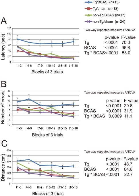
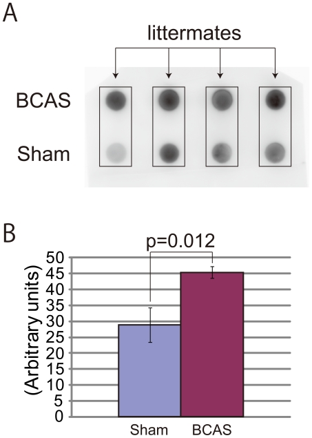
Methods: Two months old mice overexpressing a mutant form of the human APP bearing both the Swedish and Indiana mutations (APP(Sw/Ind)-Tg mice), or their wild-type littermates, were subjected to chronic cerebral hypoperfusion with bilateral common carotid artery stenosis (BCAS) using microcoils or sham operation. Barnes maze test performance and histopathological findings were analyzed at eight months old by 2 ×ばつ 2 factorial experimental designs with four groups.
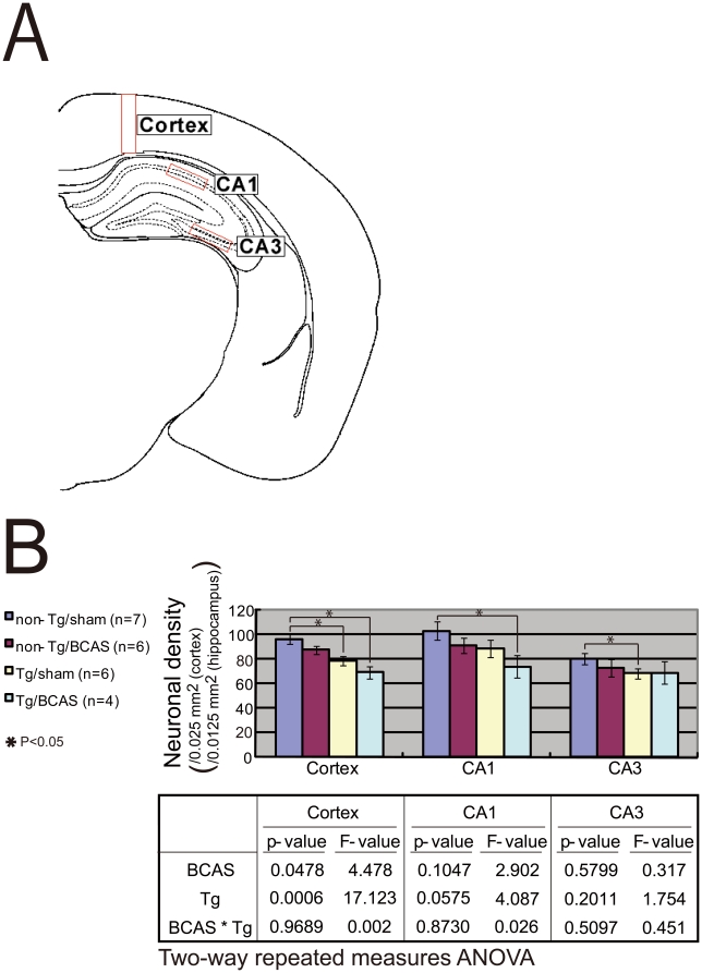
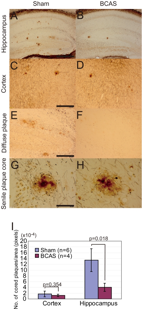
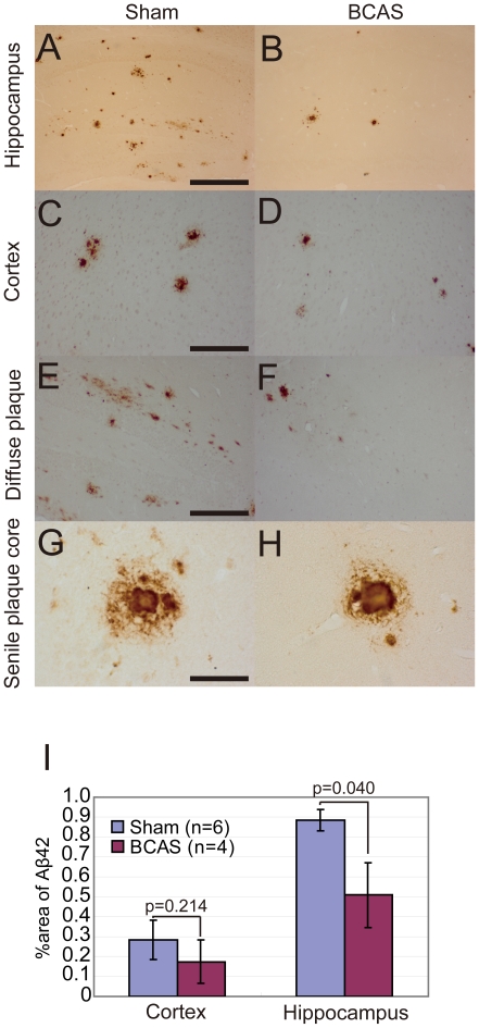
Results: BCAS-operated APP(Sw/Ind)-Tg mice showed significantly impaired learning ability compared to the other three groups of mice. Two-way repeated measures analysis of variance showed a synergistic interaction between the APP genotype and BCAS operation in inducing learning impairment. The cognitive performances were significantly correlated with the neuronal densities. BCAS significantly reduced the density of Nissl-stained neurons and silver-stained cored plaques in the hippocampus of APP(Sw/Ind)-Tg mice but increased the amount of filter-trap amyloid β in the extracellular-enriched soluble brain fraction, compared to those from sham operated mice.
Conclusions: The results suggest interaction between chronic cerebral hypoperfusion and APP(Sw/Ind) overexpression in cognitive decline in mice through enhanced neuronal loss and altered amyloid β metabolism.
Conflict of interest statement
Figures
References
-
- Hachinski V, Iadecola C, Petersen RC, Breteler MM, Nyenhuis DL, et al. National Institute of Neurological Disorders and Stroke-Canadian Stroke Network vascular cognitive impairment harmonization standards. Stroke. 2006;37:2220–2241. - PubMed
-
- Kalaria R. Similarities between Alzheimer's disease and vascular dementia. J Neurol Sci. 2002;203-204:29–34. - PubMed
-
- de la Torre JC. Alzheimer disease as a vascular disorder: nosological evidence. Stroke. 2002;33:1152–1162. - PubMed
-
- Kalaria RN. Cerebral vessels in ageing and Alzheimer's disease. Pharmacol Ther. 1996;72:193–214. - PubMed
-
- Kitaguchi H, Ihara M, Saiki H, Takahashi R, Tomimoto H. Capillary beds are decreased in Alzheimer's disease, but not in Binswanger's disease. Neurosci Lett. 2007;417:128–131. - PubMed
Publication types
MeSH terms
Substances
Grants and funding
LinkOut - more resources
Full Text Sources
Molecular Biology Databases
Miscellaneous