This site needs JavaScript to work properly. Please enable it to take advantage of the complete set of features!
Skip to main page content
U.S. flag

An official website of the United States government

Dot gov

The .gov means it’s official.
Federal government websites often end in .gov or .mil. Before sharing sensitive information, make sure you’re on a federal government site.

Https

The site is secure.
The https:// ensures that you are connecting to the official website and that any information you provide is encrypted and transmitted securely.

NIH NLM Logo
Log in
Access keys NCBI Homepage MyNCBI Homepage Main Content Main Navigation
. 2010 Oct 15;285(42):32312-24.
doi: 10.1074/jbc.M110.124917. Epub 2010 Aug 11.

Ablation of succinate production from glucose metabolism in the procyclic trypanosomes induces metabolic switches to the glycerol 3-phosphate/dihydroxyacetone phosphate shuttle and to proline metabolism

Affiliations

Ablation of succinate production from glucose metabolism in the procyclic trypanosomes induces metabolic switches to the glycerol 3-phosphate/dihydroxyacetone phosphate shuttle and to proline metabolism

Charles Ebikeme et al. J Biol Chem. .

Abstract

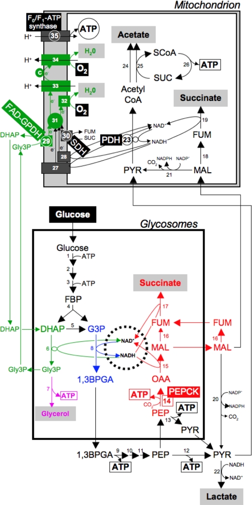
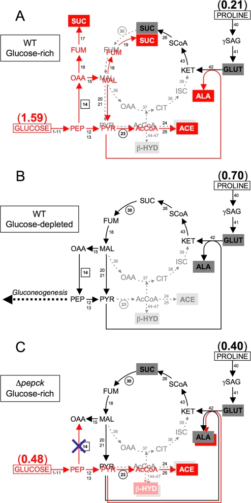
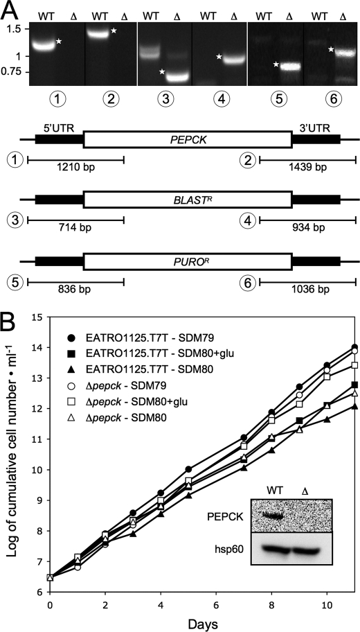
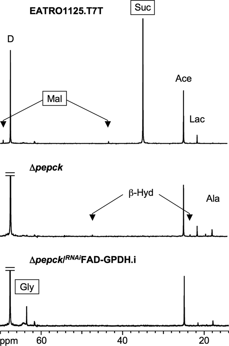
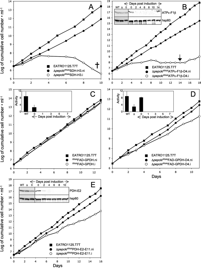
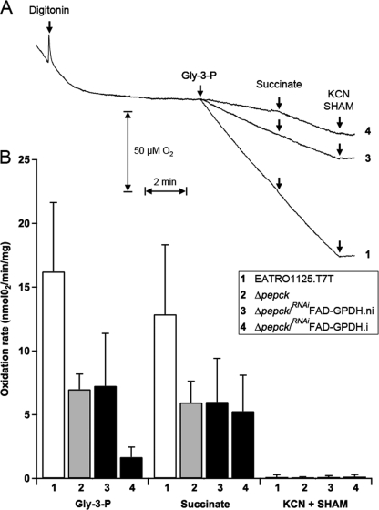
Trypanosoma brucei is a parasitic protist that undergoes a complex life cycle during transmission from its mammalian host (bloodstream forms) to the midgut of its insect vector (procyclic form). In both parasitic forms, most glycolytic steps take place within specialized peroxisomes, called glycosomes. Here, we studied metabolic adaptations in procyclic trypanosome mutants affected in their maintenance of the glycosomal redox balance. T. brucei can theoretically use three strategies to maintain the glycosomal NAD(+)/NADH balance as follows: (i) the glycosomal succinic fermentation branch; (ii) the glycerol 3-phosphate (Gly-3-P)/dihydroxyacetone phosphate (DHAP) shuttle that transfers reducing equivalents to the mitochondrion; and (iii) the glycosomal glycerol production pathway. We showed a hierarchy in the use of these glycosomal NADH-consuming pathways by determining metabolic perturbations and adaptations in single and double mutant cell lines using a combination of NMR, ion chromatography-MS/MS, and HPLC approaches. Although functional, the Gly-3-P/DHAP shuttle is primarily used when the preferred succinate fermentation pathway is abolished in the Δpepck knock-out mutant cell line. In the absence of these two pathways (Δpepck/(RNAi)FAD-GPDH.i mutant), glycerol production is used but with a 16-fold reduced glycolytic flux. In addition, the Δpepck mutant cell line shows a 3.3-fold reduced glycolytic flux compensated by an increase of proline metabolism. The inability of the Δpepck mutant to maintain a high glycolytic flux demonstrates that the Gly-3-P/DHAP shuttle is not adapted to the procyclic trypanosome context. In contrast, this shuttle was shown earlier to be the only way used by the bloodstream forms of T. brucei to sustain their high glycolytic flux.


Figures

FIGURE 1.
FIGURE 1.
Schematic representation of glucose metabolism in the procyclic form of T. brucei. This figure describes the glycosomal NADH producing and consuming pathways, highlighted by a dashed circle and colored pathways. The glycosomal NADH-producing step is shown in blue; the glycosomal succinic fermentation pathways is shown in red; the Gly-3-P/DHAP shuttle and the associated complexes of the respiratory chain are shown in green; and the glycerol-producing step is shown in purple. Excreted end products from glucose metabolism are shown in black, red, green, or purple characters on a gray rectangle as background. ATP molecules produced by substrate level phosphorylation and oxidative phosphorylation are boxed and circled, respectively. Enzymatic steps targeted by RNAi are circled, and the PEPCK step, in which the gene has been deleted, is boxed; the name of the genetically manipulated enzymes is also indicated. Abbreviations used are as follows: 1,3BPGA, 1,3-bisphosphoglycerate; c, cytochrome c; e, electrons; FBP, fructose 1,6-bisphosphate; FUM, fumarate; G3P, glyceraldehyde 3-phosphate; Gly3P, glycerol 3-phosphate; MAL, malate; OAA, oxaloacetate; PEP, phosphoenolpyruvate; PYR, pyruvate; SCoA, succinyl-CoA; SUC, succinate. Enzymes used are as follows: steps 1, hexokinase; 2, glucose-6-phosphate isomerase; 3, phosphofructokinase; 4, aldolase; 5, triose-phosphate isomerase; 6, NADH-dependent glycerol-3-phosphate dehydrogenase; 7, glycerol kinase; 8, glyceraldehyde-3-phosphate dehydrogenase; 9, phosphoglycerate kinase; 10, phosphoglycerate mutase; 11, enolase; 12, pyruvate kinase; 13, pyruvate phosphate dikinase; 14, phosphoenolpyruvate carboxykinase (PEPCK, Tb927.2.4210); 15, malate dehydrogenase; 16, cytosolic fumarase; 17, glycosomal NADH-dependent fumarate reductase; 18, mitochondrial fumarase; 19, mitochondrial NADH-dependent fumarate reductase; 20, cytosolic malic enzyme; 21, mitochondrial malic enzyme; 22, unknown enzyme; 23, pyruvate dehydrogenase complex (PDH-including the E2 subunit, PDH-E2, Tb927.10.7570); 24, unknown enzyme; 25, acetate:succinate CoA-transferase; 26, succinyl-CoA synthetase; 27, rotenone-sensitive NADH dehydrogenase (complex I of the respiratory chain); 28, rotenone-insensitive NADH dehydrogenase; 29, FAD-dependent glycerol-3-phosphate dehydrogenase (FAD-GPDH, Tb11.02.5280); 30, succinate dehydrogenase (SDH, complex II of the respiratory chain, Tb927.8.6580); 31, ubiquinone; 32, SHAM-sensitive alternative oxidase; 33, complex III of the respiratory chain; 34, complex IV of the respiratory chain; 35, F0F1-ATP synthase (ATPε- including the F1β subunit, ATPε-F1β, Tb927.3.1380).
FIGURE 2.
FIGURE 2.
Schematic representation of proline and glucose metabolism in the wild type and Δpepck procyclic trypanosomes. A and B represent the wild type cells grown in glucose-rich and glucose-depleted conditions, respectively. C describes the switch to proline metabolism observed for the Δpepck mutant grown in glucose-rich conditions. Major and minor end products from proline metabolism are black characters on dark and light gray rectangles as backgrounds, respectively. End products from glucose metabolism are white characters on red rectangles. Dashed gray arrows indicate steps, which are considered to occur at a background level or not at all. The rate of proline and/or glucose consumption (μmol consumed/h/mg of protein) is indicated by parentheses. Deletion of the PEPCK gene is represented by a blue cross in C. Enzymatic steps targeted by RNAi are circled, and the PEPCK step, in which the gene has been deleted, is boxed. Abbreviations not used in Fig. 1 are as follows: ACE, acetate; AcCoA, acetyl-CoA; ALA, alanine; β-HYD, β-hydroxybutyrate; GLUT, glutamate; γSAG, glutamate γ-semialdehyde. Enzymes not shown in Fig. 1 are as follows: steps 36, mitochondrial malate dehydrogenase; 37, citrate synthase; 38, aconitase; 39, isocitrate dehydrogenase; 40, l-proline dehydrogenase; 41, pyrroline-5-carboxylate dehydrogenase; 42, l-alanine aminotransferase; 43, 2-ketoglutarate dehydrogenase complex; 44; acetyl-CoA acetyltransferase; 45, 3-hydroxy-3-methylglutaryl-CoA synthase; 46, 3-hydroxy-3-methylglutaryl-CoA lyase; 47, β-hydroxybutyrate dehydrogenase.
FIGURE 3.
FIGURE 3.
Analysis of the Δpepck mutant cell line. A shows a PCR analysis of genomic DNA isolated from the parental EATRO1125.T7T (WT) and Δpepck (Δ) cell lines. Amplifications were performed with primers based on sequences that flank the 5′UTR and 3′UTR fragments used to target the PEPCK gene depletion (black boxes) and internal sequences from the PEPCK gene (PCR products 1 and 2), the blasticidin resistance gene (BLASTR, PCR products 3 and 4), or the puromycin resistance gene (PUROR, PCR products 5 and 6). As expected, PCR amplification using primers derived from the PEPCK gene and drug-resistant genes were only observed for the parental EATRO1125.T7T (WT) and Δpepck (Δ) cell lines, respectively (DNA bands labeled with a star). B shows the growth curve of the EATRO1125.T7T and Δpepck cell lines incubated in SDM79 and the SDM80 medium either containing (SDM80glu) or lacking (SDM80) glucose. Cells were maintained in the exponential growth phase (between 106 and 107 cells/ml), and cumulative cell numbers reflect normalization for dilution during cultivation. The inset shows a Western blot analysis of the parental (WT) and Δpepck (Δ) cell lines with the anti-PEPCK and anti-hsp60 immune sera.
FIGURE 4.
FIGURE 4.
Carbon-13 NMR spectra of metabolic end products excreted by procyclic cell lines incubated with d-[1-13C]glucose. For these NMR analyses, the parental EATRO1125.T7T, the Δpepck, and tetracycline-induced Δpepck/RNAiFAD-GPDH.i cell lines were incubated with 4 mm d-[1-13C]glucose in PBS/NaHCO3 buffer. The NMR spectra were obtained after addition of 15 μl of dioxane. Each spectrum corresponds to one representative experiment from a set of at least three. The boxed metabolites (succinate, malate, and glycerol) are excreted metabolites produced in the glycosomes. The resonances were assigned as follows: Ala, alanine; Ace, acetate; β-Hyd, β-hydroxybutyrate; D, dioxane; Gly, glycerol; Lac, lactate; Mal, malate; Suc, succinate.
FIGURE 5.
FIGURE 5.
Analysis of mutant cell lines. A–E show the growth curve of the Δpepck/RNAiSDH, Δpepck/RNAiATPε-F1β, RNAiFAD-GPDH, Δpepck/RNAiFAD-GPDH, and Δpepck/RNAiPDH-E2 cell lines, respectively, incubated in SDM79 in the presence (.i, しろまる) or in the absence (.ni, くろまる) of tetracycline. Cells were maintained in the exponential growth phase (between 106 and 107 cells/ml), and cumulative cell numbers reflect normalization for dilution during cultivation. The cross indicates that further incubation led to the death of the whole population (A), and the arrow indicates the observed reexpression of the ATPε-F1β protein (B). The insets in B and E show Western blot analyses of the EATRO1125.T7T (WT) and Δpepck (Δ) cell lines, plus the Δpepck/RNAiATPε-F1β (B) or Δpepck/RNAiPDH-E2 (E) cell lines upon tetracycline addition (lanes 0–10/14). The name of the proteins recognized by the immune sera is indicated in the right margin. The insets in C and D show the FAD-GPDH activity (milliunits/mg of protein) of the EATRO1125.T7T parental cell line (WT) plus the RNAiFAD-GPDH (C) or Δpepck/RNAiFAD-GPDH (D) cell lines upon tetracycline addition (lanes 0–7). The inset of D also shows the FAD-GPDH activity of the Δpepck mutant (Δ). The FAD-GPDH activities are normalized with the malate dehydrogenase activity measured in the same samples.
FIGURE 6.
FIGURE 6.
HPLC analysis of metabolic end products excreted by procyclic cell lines incubated with d-[1-13C]glucose. Metabolites contained in incubation medium of uninduced (A) and tetracycline-induced (B) Δpepck/RNAiFAD-GPDH cells were separated by HPLC and analyzed by refractometry. The same samples were analyzed by NMR (Table 3 and Fig. 4). Abbreviations used are as follows: Ace, acetate; Gly, glycerol; Lac, lactate; Suc, succinate; RIU, refractive index unit.
FIGURE 7.
FIGURE 7.
Oxygen consumption in permeabilized procyclic T. brucei cells. A, trace of respiration rates in 3 ×ばつ 108 EATRO1125.T7T (1), Δpepck/RNAiFAD-GPDH.ni (3), and Δpepck/RNAiFAD-GPDH.i (4) cells, upon the addition of digitonin (60 μg), Gly-3-P (12.5 mm), succinate (12.5 mm), and KCN/SHAM (6.25/1.56 mm). B, rate of oxygen consumption for the EATRO1125.T7T (1), Δpepck (2), Δpepck/RNAiFAD-GPDH.ni (3), and Δpepck/RNAiFAD-GPDH.i (4) cell lines (4 < n < 6, error bars are ± S.E.).

References

    1. Barrett M. P., Burchmore R. J., Stich A., Lazzari J. O., Frasch A. C., Cazzulo J. J., Krishna S. (2003) Lancet 362, 1469–1480 - PubMed
    1. Michels P. A., Bringaud F., Herman M., Hannaert V. (2006) Biochim. Biophys. Acta 1763, 1463–1477 - PubMed
    1. Cross G. A., Klein R. A., Linstead D. J. (1975) Parasitology 71, 311–326 - PubMed
    1. Coustou V., Besteiro S., Biran M., Diolez P., Bouchaud V., Voisin P., Michels P. A., Canioni P., Baltz T., Bringaud F. (2003) J. Biol. Chem. 278, 49625–49635 - PubMed
    1. Fairlamb A. H., Opperdoes F. R. (1986) in Carbohydrate Metabolism in Cultured Cells (Morgan M. J. ed) pp. 183–224, Plenum Publishing Corp., New York

Publication types

MeSH terms

Cite

AltStyle によって変換されたページ (->オリジナル) /