This site needs JavaScript to work properly. Please enable it to take advantage of the complete set of features!
Skip to main page content
U.S. flag

An official website of the United States government

Dot gov

The .gov means it’s official.
Federal government websites often end in .gov or .mil. Before sharing sensitive information, make sure you’re on a federal government site.

Https

The site is secure.
The https:// ensures that you are connecting to the official website and that any information you provide is encrypted and transmitted securely.

NIH NLM Logo
Log in
Access keys NCBI Homepage MyNCBI Homepage Main Content Main Navigation
. 2010 Jul 8;5(7):e11479.
doi: 10.1371/journal.pone.0011479.

Endocytosis of chikungunya virus into mammalian cells: role of clathrin and early endosomal compartments

Affiliations

Endocytosis of chikungunya virus into mammalian cells: role of clathrin and early endosomal compartments

Eric Bernard et al. PLoS One. .

Abstract

Background: The replicative cycle of chikungunya virus (CHIKV), an alphavirus that recently re-emerged in India and in Indian Ocean area, remains mostly unknown. The aim of the present study was to investigate the intracellular trafficking pathway(s) hijacked by CHIKV to enter mammalian cells.

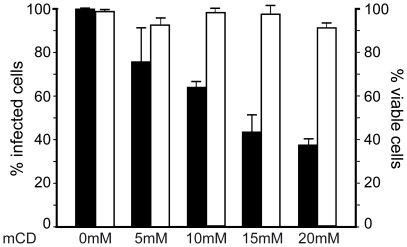
Methodology/principal findings: Entry pathways were investigated using a variety of pharmacological inhibitors or overexpression of dominant negative forms of proteins perturbating cellular endocytosis. We found that CHIKV infection of HEK293T mammalian cells is independent of clathrin heavy chain and- dependent of functional Eps15, and requires integrity of Rab5-, but not Rab7-positive endosomal compartment. Cytoskeleton integrity is crucial as cytochalasin D and nocodazole significantly reduced infection of the cells. Finally, both methyl beta-cyclodextrin and lysomotropic agents impaired CHIKV infection, supporting that a cholesterol-, pH-dependent step is required to achieve productive infection. Interestingly, differential sensitivity to lysomotropic agents was observed between the prototypal 37997 African strain of CHIKV and the LR-OPY1 virus isolated from the recent outbreak in Reunion Island.

Conclusions: Together our data indicate that CHIKV entry in its target cells is essentially mediated by clathrin-independent, Eps15-dependent endocytosis. Despite that this property is shared by the prototypal 37997 African strain of CHIKV and the LR-OPY1 virus isolated from the recent outbreak in La Réunion Island, differential sensitivity to lysomotropic agents may support that the LR-OPY1 strain has acquired specific entry mechanisms.


Conflict of interest statement

Competing Interests: The authors have declared that no competing interests exist.

Figures

Figure 1
Figure 1. Functional Eps15 but not clathrin heavy chain expression is required for infection by CHIKV.
HEK293T cells were transfected with plasmids encoding either a GFP-labelled wild type Eps15 (WT) or the GFP-tagged dominant negative Eps15Δ95/295 mutant (DN) and maintained for 36 h in culture. (A) Cells were incubated with Alexa595-labelled transferrin for 30 min at 37°C and uptake by transfected cells was monitored using immunofluorescence microscopy. (B) Transfected cells were challenged with CHIKV for 4 h at 37°C (37997-CHIKV strain used at a m.o.i. of 5) and the presence of intracellular viral antigens was detected using a mAbs C42 reacting with CHIKV and Texas-red-conjugated secondary antibodies. (C) Tansgene-expressing cells positive for intracellular CHIKV antigens were quantified from panel (B) by flow cytometry. Values are the mean of 4 separate experiments performed in triplicate ± SD. (D) Cells transfected with siRNA targeting the clathrin heavy chain (CHC) or with control siRNA (Ctrl.) were challenged with 37997-CHIKV. Levels of infection were monitored by quantification of GFP-positive cells using flow cytometry (upper panel). Levels of CHC expression in transfected cells were analyzed by immunoblot analysis. Actin expression was used to monitor proteins level in each sample (lower panel).
Figure 2
Figure 2. CHIKV infection of HEK293T cells is inhibited by lysomotropic agents.
(A) Cells were incubated in medium supplemented with drug solvent (Med) or medium supplemented with appropriate concentrations of lysomotropic agents inhibiting endosomal acidification: chloroquine (Chl), monensine (Mo), amonium chloride (NH4Cl), bafilomycine A1 (Baf). The cells were challenged with the 37997-GFP CHIKV strain used at a m.o.i. of 5. The percentage of GFP-expressing cells was determined by flow cytometry analysis (black bars). For each experiment, the number of viable cells was determined after exclusion coloration with Trypan Blue (white bars). Values are expressed as a percentage of untreated controls. Each value is the mean of 3 separate experiments performed in triplicate ± SD. (B) Appropriate concentrations of lysomotropic drugs were added to CHIKV infected cells 4 hours after viral challenge (m.o.i. of 5). After an additional 16 h in culture, percentage of infected cells was determined by flow cytometry.
Figure 3
Figure 3. Methyl β-cyclodextrin treatment decreases infection of mammalian cells by CHIKV.
HEK293T cells were incubated in medium alone or medium supplemented with increasing concentrations of methyl β-cyclodextrin (mCD) and challenged with the 37997-GFP CHIKV strain (m.o.i. of 5). The percentage of GFP expressing cells was determined by flow cytometry analysis (black histograms). For each experiment, the number of viable cells was determined after exclusion coloration with Trypan Blue (white histograms). Values are expressed as a percentage of untreated conditions. Each value is the mean of 3 separate experiments performed in triplicate ± SD.
Figure 4
Figure 4. Role of Rab5 and Rab7 GTPases in CHIKV infection of HEK293T cells.
HEK293T cells grown on coverslips were transfected with plasmids encoding either wild-type (WT) or dominant negative (DN) forms of GFP-tagged Rab5 (A), (B) and (D) or GFP-tagged Rab7 (C) and (E). Twenty-four hours post-transfection, cells were challenged with CHIKV (37997 strain used at a m.o.i. of 5). After another 4 hours in culture, the cells were permeabilized and viral antigens were labelled using anti-capsid C42 mAb and Alexa 594 secondary anti-Ig reagents. Antigen-expressing cells were revealed by confocal imaging (B) and (C), or quantified by flow cytometry (D) and (E). In control experiments, cells expressing GFP-fused WT or DN Rab5 were analyzed for Alexa Fluor 594-labelled transferrin uptake by confocal imaging (A). All experiments were repeated three times with similar outcomes. Images are representative of individual cells visualized and percentages of CHIKV positive cells determined by flow cytometry analysis are the mean of 4 separate experiments performed in triplicate ± SD.
Figure 5
Figure 5. Effect of actin-disrupting drug and microtubule depolymerizating agent on CHIKV infection.
HEK293T cells were treated with increasing concentrations of (A) cytochalasin D (Cyt D) or (B) nocodazole (NoD) before exposure to the 37997-GFP strain of CHIKV (m.o.i. of 5) (black bars). Sixteen hours post-infection, the percentage of infected cells was determined by quantification of GFP-expressing cells by flow cytometry analysis. For each point, cell viability was determined by Trypan blue exclusion and expressed as a percentage of untreated controls (white bars). Values are the mean of 3 separate experiments performed in duplicate + SD. Organization of cytoskeleton in cells cultured in medium alone (Med) or supplemented with cytochalsin D (C) or nocodazole (D) was analyzed by immunofluorescence after labelling with anti-actin (C) or anti-tubulin mAbs (D). Nuclei are stained with DAPI. (E) Flow cytometry analysis of CHIKV infected cells after treatment with cytochalasine D or nocodazole and viral challenge performed in neutral (black bars) or acid (white bars) medium. Each value is the mean of 3 separate experiments performed in duplicate ± SD.
Figure 6
Figure 6. Comparative analysis of entry pathways used by the 37997 African strain and the LR-OPY1 isolate to infect HEK293T cells.
Cells were challenged with normalized amounts (m.o.i. of 5) of the 37997-GFP (black bars) or the LR-OPY1-GFP (white bars) strains of CHIKV after (A) transfection of cells with siRNA directed to clathrin heavy chain (CHC) or control siRNA (Ctrl), or after treatment with (B) lysomotropic agents (chloroquine (Chl), monensin (Mo), amonium chloride (NH4Cl) or bafilomycin A1 (Baf)) or (C) cytochalasine D (CytD) or (D) nocodazole (NoD) in conditions described inMaterial and Methods. For each experiment, a control performed by incubating the cells in the presence of medium supplemented with an equivalent amount of drug solvent (Med) is included. Twenty-four hours post-infection, CHIKV replication was monitored by flow cytometry analysis of GFP expression. Each value is the mean of 3 separate experiments performed in triplicate ± SD.

References

    1. Karabatsos N. International Catalogue of Arboviruses Third edition 1985. - PubMed
    1. Strauss JH, Strauss EG. The alphaviruses: gene expression, replication, and evolution. Microbiol Rev. 1994;58:491–562. - PMC - PubMed
    1. Higashi N, Matsumoto A, Tabata K, Nagatomo Y. Electron microscope study of development of Chikungunya virus in green monkey kidney stable (VERO) cells. Virology. 1967;33:55–69. - PubMed
    1. Simizu B, Yamamoto K, Hashimoto K, Ogata T. Structural proteins of Chikungunya virus. J Virol. 1984;51:254–258. - PMC - PubMed
    1. Ross RW. The Newala epidemic. III. The virus: isolation, pathogenic properties and relationship to the epidemic. J Hyg (Lond) 1956;54:177–191. - PMC - PubMed

Publication types

MeSH terms

Cite

AltStyle によって変換されたページ (->オリジナル) /