This site needs JavaScript to work properly. Please enable it to take advantage of the complete set of features!
Skip to main page content
U.S. flag

An official website of the United States government

Dot gov

The .gov means it’s official.
Federal government websites often end in .gov or .mil. Before sharing sensitive information, make sure you’re on a federal government site.

Https

The site is secure.
The https:// ensures that you are connecting to the official website and that any information you provide is encrypted and transmitted securely.

NIH NLM Logo
Log in
Access keys NCBI Homepage MyNCBI Homepage Main Content Main Navigation
. 2009 Dec;5(12):e1000683.
doi: 10.1371/journal.ppat.1000683. Epub 2009 Dec 11.

The T3SS effector EspT defines a new category of invasive enteropathogenic E. coli (EPEC) which form intracellular actin pedestals

Affiliations

The T3SS effector EspT defines a new category of invasive enteropathogenic E. coli (EPEC) which form intracellular actin pedestals

Richard Bulgin et al. PLoS Pathog. 2009 Dec.

Abstract

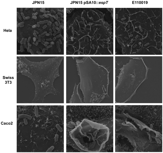
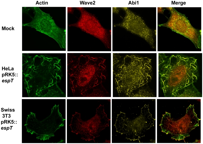
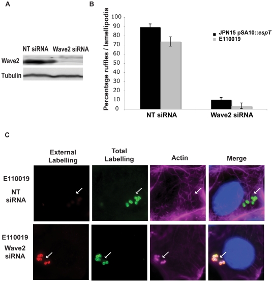
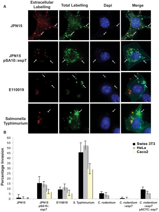
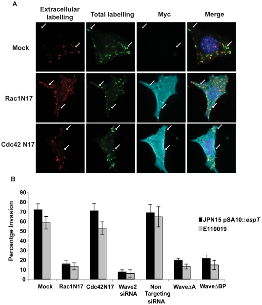
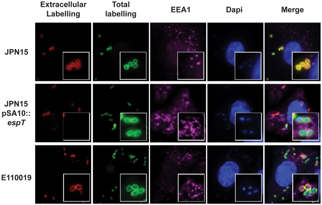
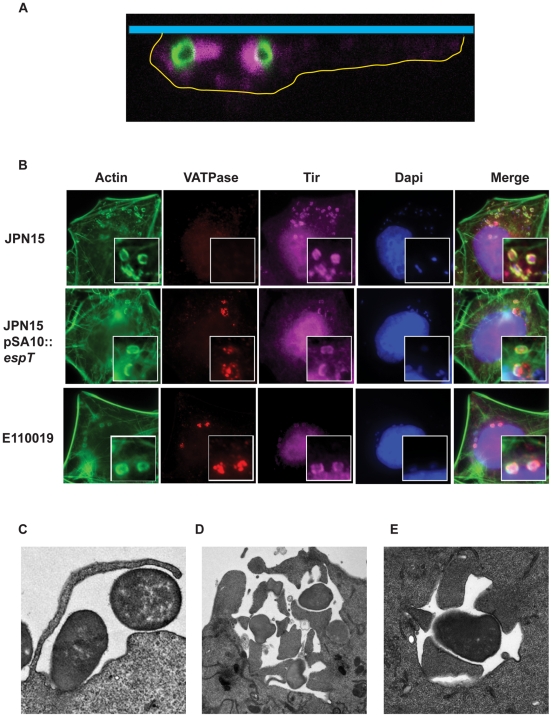
Enteropathogenic Escherichia coli (EPEC) strains are defined as extracellular pathogens which nucleate actin rich pedestal-like membrane extensions on intestinal enterocytes to which they intimately adhere. EPEC infection is mediated by type III secretion system effectors, which modulate host cell signaling. Recently we have shown that the WxxxE effector EspT activates Rac1 and Cdc42 leading to formation of membrane ruffles and lamellipodia. Here we report that EspT-induced membrane ruffles facilitate EPEC invasion into non-phagocytic cells in a process involving Rac1 and Wave2. Internalized EPEC resides within a vacuole and Tir is localized to the vacuolar membrane, resulting in actin polymerization and formation of intracellular pedestals. To the best of our knowledge this is the first time a pathogen has been shown to induce formation of actin comets across a vacuole membrane. Moreover, our data breaks the dogma of EPEC as an extracellular pathogen and defines a new category of invasive EPEC.


Conflict of interest statement

The authors have declared that no competing interests exist.

Figures

Figure 1
Figure 1. Scanning electron microscopy of HeLa, Swiss 3T3 and polarized Caco2 cells infected with JPN15, JPN15 expressing EspT or E110019 for 2 h.
JPN15 displayed a pattern of diffused adherence on HeLa, Swiss 3T3 and Caco2 cells but did not induce any significant membrane remodeling. JPN15 expressing EspT and E110019 also adhered to HeLa, Swiss 3T3 and Caco2 cells in a diffuse pattern but induced membrane ruffles at the site of bacterial attachment, which were more pronounced on Caco2 cells. In addition to membrane ruffles JPN15 expressing EspT and E110019 also induced formation of lamellipodia and dorsal ruffles at locations distal from the site of bacterial attachment on Swiss 3T3 cells. Magnifications: HeLa cells X5000; Swiss 3T3 X3500; Caco2 X10000 and X6500 (middle).
Figure 2
Figure 2. Wave2 and Abi1 are localized to membrane ruffles and lamellipodia induced by ectopically expressed EspT.
Actin was stained with Oregon Green phalloidin (Green), Wave2 was detected with a polyclonal rabbit antibody (Red) and Abi1 was visualized using a mouse monoclonal antibody (Yellow). Mock transfected cells did not display any significant actin structures and Wave2 and Abi1 were localized diffusely in the cytoplasm. HeLa cells transfected with pRK5::espT exhibit prominent membrane ruffles on their apical surface which were enriched with Wave2 and Abi1. Ectopic expression of EspT in Swiss 3T3 cells resulted in formation of distinctive lamellipodia to which Wave2 and Abi1 were extensively recruited and co-localized.
Figure 3
Figure 3. Wave2 is essential for EspT induced membrane remodeling and invasion.
(A) Swiss 3T3 cells were treated with Non Targeting (NT) siRNA or siRNA targeted against Wave2. (A) Western blot with lysates from Swiss 3T3 cells treated with NT and Wave2 siRNA. Wave2 and Tubulin were detected with monoclonal antibodies. Non Targeting siRNA did not alter Wave2 expression whereas treatment with Wave2 siRNA depleted the protein. Protein levels in the lysates were normalized using anti tubulin antibodies. (B) Quantification of membrane remodeling induced by E110019 and JPN15 expressing EspT in Swiss 3T3 cells treated with NT and Wave2 siRNA after 3 h of infection. 100 cells were counted in triplicate resulted are presented as mean±SEM. Treatment with Wave2 siRNA oligos reduced the level of membrane remodeling induced by JPN15 pSA10::espT and E110019 to 10% and 5% respectively. This is comparable to the 9% of cells which display membrane ruffle and lamellipodia formation in cells infected with EPEC not expressing EspT. (C) Cells were infected with E110019 for 3 h were fixed and stained prior to permeabilization (extracellular labeling) (Red). The cells were then washed, permeabilized, re-labeled (Total labeling) (Green) along with Alexaflour 633 Phalloidin (Cyan) and Dapi (Blue). In cells treated with the NT siRNA E110019 induced formation of membrane ruffles and a large proportion of bacteria were labeled by the total stain which were absent from the extracellular labeling. Depletion of Wave2 using siRNA inhibited formation of membrane ruffles by E110019 and the majority of bacteria were detected by both the extracellular and total staining demonstrating that depletion of Wave2 inhibits EPEC invasion.
Figure 4
Figure 4. EspT-dependent actin remodeling facilitates bacterial invasion of epithelial cells.
(A) Swiss 3T3 cells infected with JPN15, JPN15 expressing EspT, E110019 or S. Typhimurium for 3 h were fixed and stained prior to permeabilization (extracellular labeling) (Red). The cells were then washed, permeabilized, re-labeled (Total labeling) (Green) along with Alexaflour 633 Phalloidin (Cyan) and Dapi (Blue). In cells infected with JPN15 all the bacteria labeled by the total staining were also detected with the extracellular probe indicating that there was no significant invasion (highlighted with arrows). Significant numbers of bacteria labeled with the total stain, which were not represented in the extracellular staining, were seen in cells infected JPN15 expressing EspT, E110019 and S. Typhimurium indicating that there was a significant degree of bacterial invasion (highlighted with arrows). (B) Gentamycin protection assay of Swiss 3T3 and HeLa cells infected JPN15, JPN15 expressing EspT, E110019, S. Typhimurium, C. rodentium, C. rodentium ΔespT or complemented C. rodentium ΔespT and polarized Caco2 cells infected with JPN15, JPN15 expressing EspT, E110019 or S. Typhimurium. Results are representative of 3 independent experiments carried out in duplicate and are displayed as mean±SEM.
Figure 5
Figure 5. Rac1 and Wave2 are essential for EspT-mediated bacterial invasion.
(A) Swiss 3T3 cells were left untransfected or transfected with Non Targeting (NT) or Wave2 siRNA olgos, the ectopic expression vector pDSRed encoding Wave2ΔA, Wave2ΔBP or pRK5 expressing dominant negative Rac1 and Cdc42 and infected with JPN15, JPN15 expressing EspT or E110019. Mock transfected cells or cells transfected with dominant negative Cdc42 were efficiently invaded by both JPN15 expressing EspT and E110019. Cells transfected with dominant negative Rac1 were significantly more resistant to bacterial invasion. The Wave2ΔA and Wave2ΔBP constructs also had a potent dominant negative effect on bacterial internalization. (B) Quantification of bacterial invasion; cells which had 3 or more internalized bacteria were scored as invaded. 100 cells were counted in triplicate in three independent experiments. Results are displayed as mean±SEM.
Figure 6
Figure 6. Internalized EPEC bacteria are enclosed within a vacuole.
HeLa cells were infected with JPN15, JPN15 expressing EspT and E110019 for 45 min. Internalized bacteria bound within a vacuole were detected by anti EEA1 and a secondary antibody coupled to a CY5 (magenta). DNA was detected using Dapi staining. Extracellular JPN15 bacteria, which were detected by both the total stain and the extra-cellular probe, were not co-localized with EEA1. Intracellular JPN15 expressing EspT and E110019 which were detected by the total bacterial probe but not by the pre permeabilization stain were co localized within EEA1 rich structures indicative of early endosome like vacuoles.
Figure 7
Figure 7. Internalized EPEC bacteria incorporate Tir into the vacuolar membrane which nucleates intracellular actin pedestals.
(A) Swiss 3T3 cells were infected with E110019 and processed for immuno-fluorescence confocal microscopy. A series of confocal X-stacks were taken through the infected cells. The cell boundaries were defined based on actin staining and are marked by a yellow line, the coverslip is represented by the blue bar. The staining shows intracellular E110019 (Green) associated with actin pedestal-like structures (Magenta). (B) HeLa cells were infected for 60 min with JPN15, JPN15 expressing EspT, and E110019 were processed for immuno-fluorescence microscopy after 8 h gentamycin treatment. Actin was stained using Oregon Green phalloidin (Green), Tir was detected using polyclonal Tir antisera (Magenta), Vacuolar ATPase (VATPase) was detected using a monoclonal antibody (Red) and bacteria were detected using Dapi staining. Cells infected JPN15 recruited Tir to the site of bacterial attachment and form canonical actin rich pedestals but do not display any bacterial co-localization with the VATPase vacuolar marker. In cells infected with JPN15 expressing EspT or E110019 a proportion of bacteria were co-localized with VATPase, Tir and actin. (C) A TEM micrograph showing membrane ruffles engulfing E110019. (D) E110019 has the capacity to form multiple pedestals during ruffle formation and closure. (E) E110019 bacteria bound within a vacuole with intracellular pedestals formed around its circumference.

References

    1. Nataro JP, Kaper JB. Diarrheagenic Escherichia coli. Clin Microbiol Rev. 1998;11:142–201. - PMC - PubMed
    1. Mundy R, MacDonald TT, Dougan G, Frankel G, Wiles S. Citrobacter rodentium of mice and man. Cell Microbiol. 2005;7:1697–1706. - PubMed
    1. Frankel G, Phillips AD. Attaching effacing Escherichia coli and paradigms of Tir-triggered actin polymerization: getting off the pedestal. Cell Microbiol. 2008;10:549–556. - PubMed
    1. Knutton S, Lloyd DR, McNeish AS. Adhesion of enteropathogenic Escherichia coli to human intestinal enterocytes and cultured human intestinal mucosa. Infect Immun. 1987;55:69–77. - PMC - PubMed
    1. Frankel G, Phillips AD, Rosenshine I, Dougan G, Kaper JB, et al. Enteropathogenic and enterohaemorrhagic Escherichia coli: more subversive elements. Mol Microbiol. 1998;30:911–921. - PubMed

Publication types

MeSH terms

Cite

AltStyle によって変換されたページ (->オリジナル) /