This site needs JavaScript to work properly. Please enable it to take advantage of the complete set of features!
Skip to main page content
U.S. flag

An official website of the United States government

Dot gov

The .gov means it’s official.
Federal government websites often end in .gov or .mil. Before sharing sensitive information, make sure you’re on a federal government site.

Https

The site is secure.
The https:// ensures that you are connecting to the official website and that any information you provide is encrypted and transmitted securely.

NIH NLM Logo
Log in
Access keys NCBI Homepage MyNCBI Homepage Main Content Main Navigation
Comparative Study
. 2009 Aug;31(8):432-9.
doi: 10.1111/j.1365-3024.2009.01125.x.

Recruitment of CD8(+) T cells expressing granzyme A is associated with lesion progression in human cutaneous leishmaniasis

Affiliations
Comparative Study

Recruitment of CD8(+) T cells expressing granzyme A is associated with lesion progression in human cutaneous leishmaniasis

D R Faria et al. Parasite Immunol. 2009 Aug.

Abstract

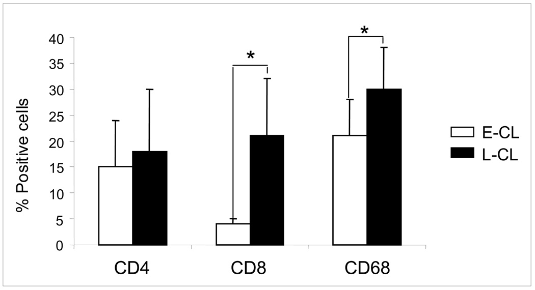
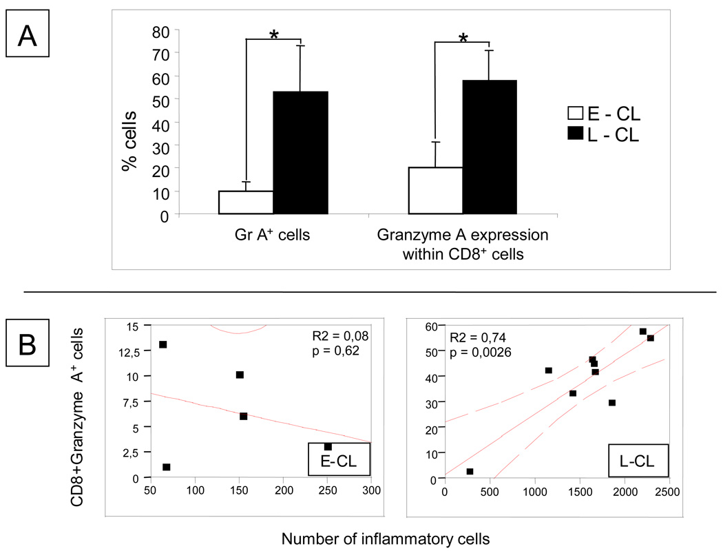
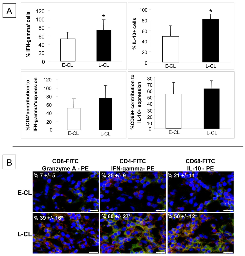
Human infection with Leishmania braziliensis leads to the establishment of cutaneous leishmaniasis (CL), characterized by the appearance of skin lesions that progress from nonulcerated to ulcerated forms. Our goal was to characterize the immunological kinetics associated with this progression, comparing the cellular composition, cytokines and granzyme expression between lesions of patients with early (E-CL) and late stages (L-CL) of CL. Histopathological analysis showed that lesions from L-CL had more exuberant inflammatory infiltrate as compared to E-CL. Although E-CL and L-CL lesions were predominantly mononuclear, lesions from E-CL patients presented higher neutrophil and eosinophil counts than L-CL. While percentages of CD4(+) and of CD68(+) cells were slightly higher in L-CL, a fivefold increase of CD8(+) cells was observed in L-CL, as compared to E-CL. Moreover, CD8(+) T-cells from L-CL expressed significantly higher levels of granzyme A than E-CL. Interestingly, granzyme A expression was positively correlated with intensity of the inflammatory infiltrate in L-CL but not E-CL. Lastly, percentages of IFN-gamma(+) and IL-10(+) cells were higher in L-CL as compared to E-CL, with CD4(+) T-cells and CD68(+) monocytes as the main sources of these cytokines, respectively. These results suggest that recruitment of CD8(+) granzyme A(+) T cells is involved in lesion progression in human CL.


Figures

Figure 1
Figure 1
Percentages of T CD4+, T CD8+ and CD68+ cells in the inflammatory infiltrate from early cutaneous leishmaniasis (E-CL) (n=6) and late cutaneous leishmaniasis (L-CL) (n=9) lesions. Frozen tissue sections were stained with FITC-labeled anti-CD4, anti-CD8 or anti-CD68 monoclonal antibodies and were counterstained with DAPI as described in Materials and Methods. Results are expressed as bars of the mean percentages for each group. Standard deviation indicated by above line bars. Asterisks indicate statistically significant differences between groups at a p value of <0.05.
Figure 2
Figure 2
(A) Percentages of cells expressing granzyme A (Gr A+) and % granzyme A expression within CD8+ T cells in early cutaneous leishmaniasis (E-CL) (n=6) and late cutaneous leishmaniasis (n= 9) lesions. (B) Correlation analysis between the frequency of CD8+granzyme A+ cells and intensity of inflammatory infiltrate in lesions from E-CL and L-CL
Figure 3
Figure 3
(A) Percentages, in early cutaneous leishmaniasis (E-CL) (n=6) and late cutaneous leishmaniasis (n= 9) lesions, of cells expressing IFN-gamma and IL-10 and the percentage contribution of CD4+ and CD68+ cells for the expression of IFN-gamma and IL-10, respectively. Frozen tissue sections were stained with FITC-labeled anti-CD4 or anti-CD68 monoclonal antibodies and with PE-labeled anti-IFN-gamma or anti-IL-10 antibody, and were counterstained with DAPI as described in Materials and Methods. Standard deviation indicated by above line bars. Asterisks indicate statistically significant differences between groups at a p value of <0.05 (t test). (B) Representative images from confocal microscopy analyses for determination of the frequencies of CD8+granzyme A, CD4+IFN-gamma+ and CD68+IL-10+ cells in E-CL and L-CL lesions. Frozen tissue sections were co-stained with FITC-labeled anti-CD8 or anti-CD4 or anti-CD68 monoclonal antibody and PE-labeled anti-granzyme A or anti-IFN-gamma or anti-IL-10, and were counterstained with DAPI as described in Materials and Methods. The three optical sections for each patient were obtained simultaneously with 363, 488 and 568 line of the argon/krypton laser and the proper set of filters. The overlay for CD4 or CD8 or CD68 (green), granzyme A or IFN-gamma or IL-10 (red) and DAPI (blue) in E-CL and L-CL lesions is shown. The cells that are double positive for each pair of staining (CD8+GranzymeA+ or CD4+IFN-gamma+ or CD8+IL-10+) appear in yellow. These images are representative of each group. Values represent the average +/− standard deviation of each group following numeric determination of the percentage of positive cells for the indicated molecules. * indicates statistically significant differences at a p value of <0.05. The bar = 10 μm.

References

    1. Schriefer A, Schriefer AL, Góes-Neto A, et al. Multiclonal Leishmania braziliensis population structure and its clinical implication in a region of endemicity for American tegumentary leishmaniasis. Infect Immun. 2004;72:508–514. - PMC - PubMed
    1. Bittencourt AL, Barral A. Evaluation of the histopathological classifications of American cutaneous and mucocutaneous leishmaniasis. Mem Inst Oswaldo Cruz. 1991;86:51–56. - PubMed
    1. Lee SA, Hasburn R. Therapy of cutaneous leishmaniasis. Int J Infect Dis. 2003;7:86–93. - PubMed
    1. Machado P, Araújo C, Da Silva AT, et al. Failure of early treatment of cutaneous leishmaniasis in preventing the development of an ulcer. Clin Infect Dis. 2002;34:69–73. - PubMed
    1. Bottrel RL, Dutra WO, Martins FA, et al. Flow cytometric determination of cellular sources and frequencies of key cytokine-producing lymphocytes directed against recombinant LACK and soluble Leishmania antigen in human cutaneous leishmaniasis. Infect Immun. 2001;69:3232–3239. - PMC - PubMed

Publication types

MeSH terms

Cite

AltStyle によって変換されたページ (->オリジナル) /