Phosphoproteomic analysis of the human pathogen Trypanosoma cruzi at the epimastigote stage
- PMID: 19579231
- PMCID: PMC2752742
- DOI: 10.1002/pmic.200800874
Phosphoproteomic analysis of the human pathogen Trypanosoma cruzi at the epimastigote stage
Abstract
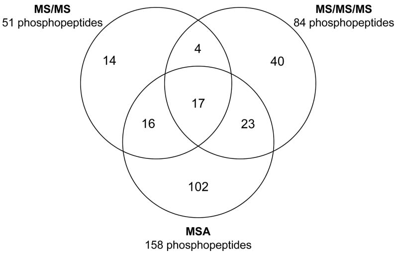
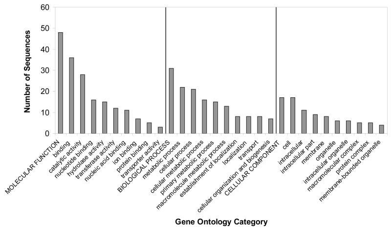
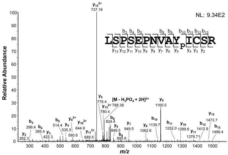
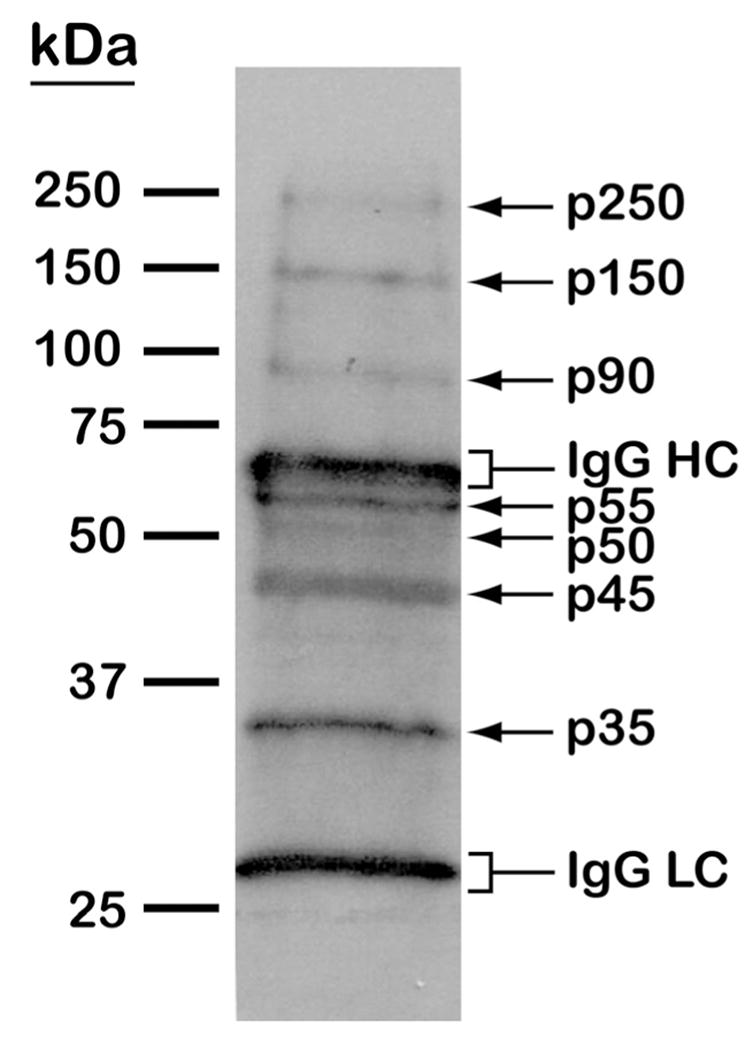
Trypanosoma cruzi is the etiologic agent of Chagas disease, which affects millions of people in Latin America and has become a public health concern in the United States and areas of Europe. The possibility that kinase inhibitors represent novel anti-parasitic agents is currently being explored. However, fundamental understanding of the cell-signaling networks requires the detailed analysis of the involved phosphorylated proteins. Here, we have performed a comprehensive MS-based phosphorylation mapping of phosphoproteins from T. cruzi epimastigote forms. Our LC-MS/MS, dual-stage fragmentation, and multistage activation analysis has identified 237 phosphopeptides from 119 distinct proteins. Furthermore, 220 phosphorylation sites were unambiguously mapped: 148 on serine, 57 on threonine, and 8 on tyrosine. In addition, immunoprecipitation and Western blotting analysis confirmed the presence of at least seven tyrosine-phosphorylated proteins in T. cruzi. The identified phosphoproteins were subjected to Gene Ontology, InterPro, and BLAST analysis, and categorized based on their role in cell structure, motility, transportation, metabolism, pathogenesis, DNA/RNA/protein turnover, and signaling. Taken together, our phosphoproteomic data provide new insights into the molecular mechanisms governed by protein kinases and phosphatases in T. cruzi. We discuss the potential roles of the identified phosphoproteins in parasite physiology and drug development.
Conflict of interest statement
The authors have declared no conflict of interest.
Figures
References
-
- Barrett MP, Burchmore RJ, Stich A, Lazzari JO, et al. The trypanosomiases. Lancet. 2003;362:1469–1480. - PubMed
-
- Dias JC, Silveira AC, Schofield CJ. The impact of Chagas disease control in Latin America: a review. Mem Inst Oswaldo Cruz. 2002;97:603–612. - PubMed
-
- Bern C, Montgomery SP, Herwaldt BL, Rassi A, Jr, et al. Evaluation and treatment of Chagas disease in the United States: a systematic review. JAMA. 2007;298:2171–2181. - PubMed
-
- Piron M, Verges M, Munoz J, Casamitjana N, et al. Seroprevalence of Trypanosoma cruzi infection in at-risk blood donors in Catalonia (Spain) Transfusion. 2008;48:1862–1868. - PubMed
-
- Tyler KM, Engman DM. The life cycle of Trypanosoma cruzi revisited. Int J Parasitol. 2001;31:472–481. - PubMed
Publication types
MeSH terms
Substances
Grants and funding
LinkOut - more resources
Full Text Sources
Research Materials