This site needs JavaScript to work properly. Please enable it to take advantage of the complete set of features!
Skip to main page content
U.S. flag

An official website of the United States government

Dot gov

The .gov means it’s official.
Federal government websites often end in .gov or .mil. Before sharing sensitive information, make sure you’re on a federal government site.

Https

The site is secure.
The https:// ensures that you are connecting to the official website and that any information you provide is encrypted and transmitted securely.

NIH NLM Logo
Log in
Access keys NCBI Homepage MyNCBI Homepage Main Content Main Navigation
. 2009 Mar;10(3):285-99.
doi: 10.1111/j.1600-0854.2008.00864.x. Epub 2008 Dec 4.

Role of the Plasmodium export element in trafficking parasite proteins to the infected erythrocyte

Affiliations

Role of the Plasmodium export element in trafficking parasite proteins to the infected erythrocyte

Justin A Boddey et al. Traffic. 2009 Mar.

Abstract

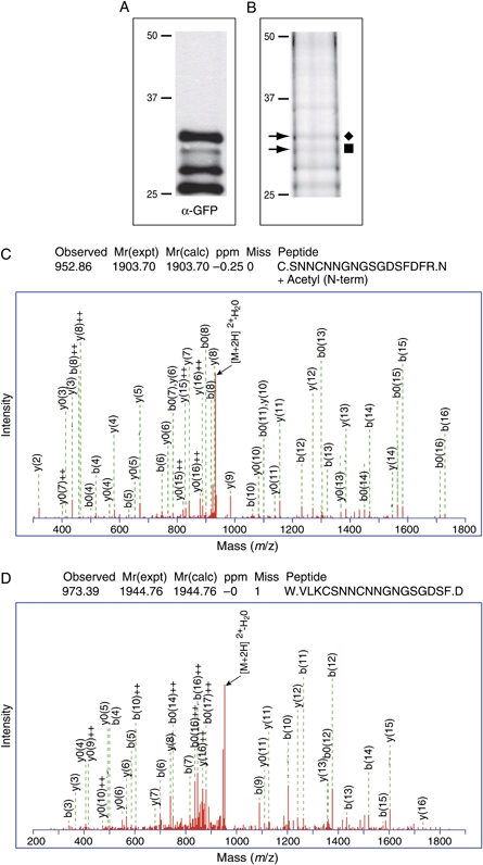
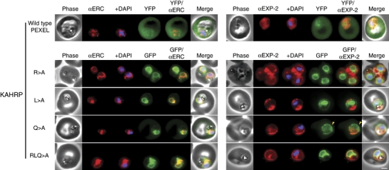
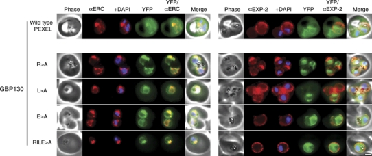
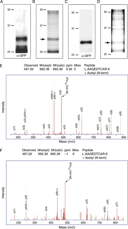
The intracellular survival of Plasmodium falciparum within human erythrocytes is dependent on export of parasite proteins that remodel the host cell. Most exported proteins require a conserved motif (RxLxE/Q/D), termed the Plasmodium export element (PEXEL) or vacuolar targeting sequence (VTS), for targeting beyond the parasitophorous vacuole membrane and into the host cell; however, the precise role of this motif in export is poorly defined. We used transgenic P. falciparum expressing chimeric proteins to investigate the function of the PEXEL motif for export. The PEXEL constitutes a bifunctional export motif comprising a protease recognition sequence that is cleaved, in the endoplasmic reticulum, from proteins destined for export, in a PEXEL arginine- and leucine-dependent manner. Following processing, the remaining conserved PEXEL residue is required to direct the mature protein to the host cell. Furthermore, we demonstrate that N acetylation of proteins following N-terminal processing is a PEXEL-independent process that is insufficient for correct export to the host cell. This work defines the role of each residue in the PEXEL for export into the P. falciparum-infected erythrocyte.


Figures

Figure 1
Figure 1. Structure of the chimaeric proteins
The first 69 residues of KAHRP or 99 residues of GBP130, containing a native or mutated PEXEL,were fused to GFP or YFP. All GBP130 chimaeras and the KAHRPWT chimaera were expressed from the HSP86 promoter . The remaining KAHRP chimaeras were expressed from the PfCRT promoter as in-frame fusions with GFPmut2.
Figure 2
Figure 2. Role of the PEXEL residues in processing and export to the erythrocyte
Immunoblots with α-GFP antibodies against KAHRP chimaeras from tetanolysin pellets (A) and supernatant (B) or GBP130 chimaeras from tetanolysin pellets (C) and supernatant (D) are shown. Antibodies to aldolase were used as a permeabilisation control, as described previously and reflects the quantity of protein loaded in each lane. While equal proportions of pellet and supernatant were loaded for each chimaera, equal loadings could not be achieved between different chimaeras because of differences in episomal expression. The densitometric analyses below (see G and H) were thus limited to comparing only between pellet and supernatant fractions of the same chimaera. Upper bands in (D, lanes 2 and 3) represent slight vacuolar leakage. E) Predicted protein sizes of KAHRP chimaeras after differential N-terminal processing; ♦ represents full-length chimaeras with signal sequence; ▪ represents processing at/near the site predicted by SignalP; H represents chimaeras processed downstream of prediction by SignalP (i.e. within the PEXEL); ▴ represents degradation to GFP/YFP only (confirmed by MS; Figure S2). The linker upstream of YFP in KAHRPWT (Figure 1) is not depicted here. While the N-termini of each KAHRP chimaera are the same, except for the mutations shown, the C-terminal YFP linker in KAHRPWT explains the minor size shift in (A) and (B) between WT and other chimaeras processed at the PEXEL (i.e. those depicted with *). F) Predicted protein sizes of GBP130 chimaeras after differential N-terminal processing. Varying exposures within the linear range of the blots represented in (A–D) were scanned at high resolution and densitometry was undertaken to approximately quantify the differential N-terminal processing and cellular localisation of KAHRP (G) and GBP130 (H) chimaeras. For each chimaera, percentages were calculated by dividing the intensity of each band in the supernatant (exported to host) or pellet (ER or parasitophorous vacuole) by the sum of the band intensities for that chimaera (total tagged chimaera) and multiplying by 100. Percentages between chimaerae are directly comparable. WT, wild type; SS, signal sequence; PV, parasitophorous vacuole.
Figure 3
Figure 3. Affinity purification of exported GBP130WT and KAHRPWT chimaeras and MS
Immunoblot(A) and coomassie gel (B) of the GBP130WT chimaera after affinity purification from the saponin supernatant. Immunoblot (C) and coomassie gel (D) of the KAHRPWT chimaera after affinity purification from the saponin supernatant. The bands indicated by an arrow in (B) and (D) were excised and subjected to MS. The lowest band (∼26 kDa) is degraded protein (YFP only). E) Mass spectra of the most N-terminal peptide from the band in (B) showing that exported GBP130 was processed in the PEXEL after leucine and acetylated at the new N-terminus.
Figure 4
Figure 4. Processing within the PEXEL occurs before export to the parasitophorous vacuole
Immunoblot with α-GFP antibodies against GBP130 chimaeras fractionated with saponin indicates that processing occurred in the parasite fraction (pellet) before export. Antibodies to aldolase were used to validate parasite membrane integrity following saponin treatment (note the presence of aldolase in the pellet fractions but absence from the supernatant fractions) and reflects the quantity of protein loaded in each lane. More of the GBP130R>A and GBP130L>A chimaera samples were loaded because of rapid loss of fluorescence over time in parasites overexpressing these mutant chimaeras. The GBP130RILE>A chimaera traffics predominantly to the parasitophorous vacuole (see supernatant fraction). That unprocessed bands of GBP130R>A, GBP130L>A and GBP130RILE>A are present in the supernatant fraction confirms breakdown of the parasitophorous vacuole membrane by saponin.
Figure 5
Figure 5. Preventing KAHRP PEXEL processing by mutation affects signal sequence processing
Immunoblot(A) and coomassie gel (B) of KAHRPR>A chimeric proteins after immunoaffinity purification from the saponin pellet. The bands indicated by arrows in (B) were excised and subjected to MS. C) Mass spectra of the most N-terminal peptide from the second upper band (▪) in (B) showing that the chimaera is processed at the site predicted by SignalP (32LKC-SNN36) and N acetylated. D) Mass spectra of the most N-terminal peptide from the uppermost band (♦) in (B) showing that the chimaera contains residues present in the signal sequence (i.e. N-terminal to the SignalP processing site; 31VLKC34) and has retained the signal sequence.
Figure 6
Figure 6. KAHRP chimaeras localise to the endoplasmic reticulum in addition to the parasitophorous vacuole or erythrocyte cytosol
Images captured by immunofluorescence microscopy show substantial colocalisation of KAHRP chimaeras with the endoplasmic reticulum protein ERC (left panels). Intraparasitic fluorescence is also evident when chimaeras were colocalised with the parasitophorous vacuole membrane protein EXP-2 (right panels). Only the wild type (WT) PEXEL chimaera (uppermost panels) is efficiently exported to the erythrocyte cytosol, but low-level fluorescence in the erythrocyte cytosol was observed for the KAHRPQ>A chimaera. All mutants show some accumulation within the parasitophorous vacuole but while the KAHRPQ>A chimaera colocalises with ERC less than the other KAHRP mutants (left panel), it accumulates more in the parasitophorous vacuole in slightly older parasites (right panel). Accumulation of the KAHRPQ>A chimaera in the parasitophorous vacuole appeared inverse to the ‘necklace of beads’ observation described previously (; see yellow arrows in right panel). The white bar in the last panel corresponds to 2 μm for each of the panels within the figure.
Figure 7
Figure 7. GBP130 chimaeras localise to the endoplasmic reticulum in addition to the parasitophorous vacuole or erythrocyte cytosol
Images captured by immunofluorescence microscopy show substantial colocalisation of GBP130 chimaeras with the endoplasmic reticulum protein ERC (left panels). Intraparasitic fluorescence is also evident when chimaeras were colocalised with the parasitophorous vacuole membrane protein EXP-2 (right panels). Only the wild-type PEXEL chimaera is efficiently exported to the erythrocyte cytosol (uppermost panels). All mutants show some accumulation within the parasitophorous vacuole and low-level fluorescence in the erythrocyte cytosol (lower panels), and this is evident, upon careful examination, in previously published images of these chimaeras . The presence of GBP130RILE>A in the saponin supernatant in Figure 4 suggests localisation predominantly in the parasitophorous vacuole (signal sequence processed). This is confirmed by immunofluorescence microscopy (compare GBP130RILE>A in this figure to KAHRPRLQ>A in Figure 6, the latter of which retains the signal sequence and localises more in the ER as a result). The white bar in the last panel corresponds to 2 μm for each of the panels within the figure.
Figure 8
Figure 8. PEXEL processing and N acetylation does not require the full-length PEXEL
Immunoblot (A) and coomassie gel (B) of the GBP130E>A chimaera after affinity purification from the saponin pellet. Immunoblot (C) and coomassie gel (D) of the GBP130E>A chimaera after affinity purification from the saponin supernatant. The bands indicated by an arrow in (B) and (D) were excised and subjected to MS. The lowest band (∼26 kDa) is degraded protein (YFP only). E) Mass spectra of the most N-terminal peptide from the band in (B) showing that GBP130E>A was processed in the parasite (pellet) within the PEXEL after leucine and N acetylated. F) Mass spectra of the most N-terminal peptide from the band in (D) showing that GBP130E>A in the parasitophorous vacuole (supernatant) was processed in the PEXEL after leucine and N acetylated.
Figure 9
Figure 9. Role of the PEXEL in export of Plasmodium falciparum proteins to the infected erythrocyte
Two proposedmodels of PEXEL-mediated export are shown (A, 1 and 2). A1) After cotranslational insertion through Sec61 at the rough endoplasmic reticulum (rER) using the signal sequence, proteins to be exported are either processed by signal peptidase (red pac-man) or sequestered and/or processed by the PEXEL protease (yellow pac-man) and sorted at the transitional endoplasmic reticulum (tER) for transport through the Golgi to the parasitophorous vacuole by the default secretory pathway. There, proteins to be exported (xE/Q/D after PEXEL processing; green protein), are recognised and trafficked across the parasitophorous vacuole membrane by a translocon. Secreted or mutated PEXEL proteins are depicted as red proteins. A2) After entry at the rER and sequestration and/or processing by either signal peptidase (red pac-man) or the PEXEL protease (yellow pac-man), proteins to be exported are differentially sorted either at the tER or at the Golgi into vesicles. This may occur through a specific transmembrane PEXEL cargo receptor that enriches functionally distinct vesicles for exported proteins (green proteins), which are targeted to subcompartments (the ‘necklace of beads’; depicted as white compartments in the parasitophorous vacuole) of the parasitophorous vacuole that houses the translocon. Exported transmembrane proteins then presumably diffuse laterally from the translocon and traffic with forming Maurer's clefts (white structures in the erythrocyte). Secreted proteins (red) traffic through the default secretory pathway to alternative compartments of the parasitophorous vacuole that do not contain the translocon. The default pathway may involve bulk flow, depicted as free red proteins in the ER that ‘sample’ the budding membrane. Uncharacterised vesicles are depicted by ‘?’. Close up (box) of the possible sorting mechanism is shown in (B) and (C). B) After cotranslational insertion into the rER PEXEL proteins are sequestered/processed by the PEXEL protease and sorted into vesicles through a transmembrane cargo receptor that interacts with the COPII machinery. C) Secreted proteins are not recognised by the PEXEL receptor but bind either a different receptor or traffic through bulk flow. The role of the Golgi is unclear but similar sorting may occur there.

References

    1. Snow RW, Guerra CA, Noor AM, Myint HY, Hay SI. The global distribution of clinical episodes of Plasmodium falciparum malaria. Nature. 2005;434:214–217. - PMC - PubMed
    1. Marti M, Baum J, Rug M, Tilley L, Cowman AF. Signal-mediated export of proteins from the malaria parasite to the host erythrocyte. J Cell Biol. 2005;171:587–592. - PMC - PubMed
    1. Smith JD, Chitnis CE, Craig AG, Roberts DJ, Hudson-Taylor DE, Peterson DS, Pinches R, Newbold CI, Miller LH. Switches in expression of Plasmodium falciparum var genes correlate with changes in antigenic and cytoadherent phenotypes of infected erythrocytes. Cell. 1995;82:101–110. - PMC - PubMed
    1. Baruch DI, Pasloske BL, Singh HB, Bi X, Ma XC, Feldman M, Taraschi TF, Howard RJ. Cloning the P.falciparum gene encoding PfEMP1, a malarial variant antigen and adherence receptor on the surface of parasitized human erythrocytes. Cell. 1995;82:77–87. - PubMed
    1. Su X-Z, Kirkman LA, Fujioka H, Wellems TE. Complex polymorphisms in an ∼330 kDa protein are linked to chloroquine-resistant P. falciparum in Southeast Asia and Africa. Cell. 1997;91:593–603. - PubMed

Publication types

Substances

LinkOut - more resources

Cite

AltStyle によって変換されたページ (->オリジナル) /