This site needs JavaScript to work properly. Please enable it to take advantage of the complete set of features!
Skip to main page content
U.S. flag

An official website of the United States government

Dot gov

The .gov means it’s official.
Federal government websites often end in .gov or .mil. Before sharing sensitive information, make sure you’re on a federal government site.

Https

The site is secure.
The https:// ensures that you are connecting to the official website and that any information you provide is encrypted and transmitted securely.

NIH NLM Logo
Log in
Access keys NCBI Homepage MyNCBI Homepage Main Content Main Navigation
. 2008 Sep 5;4(9):e1000147.
doi: 10.1371/journal.ppat.1000147.

The cysteine-rich interdomain region from the highly variable plasmodium falciparum erythrocyte membrane protein-1 exhibits a conserved structure

Affiliations

The cysteine-rich interdomain region from the highly variable plasmodium falciparum erythrocyte membrane protein-1 exhibits a conserved structure

Michael M Klein et al. PLoS Pathog. .

Abstract

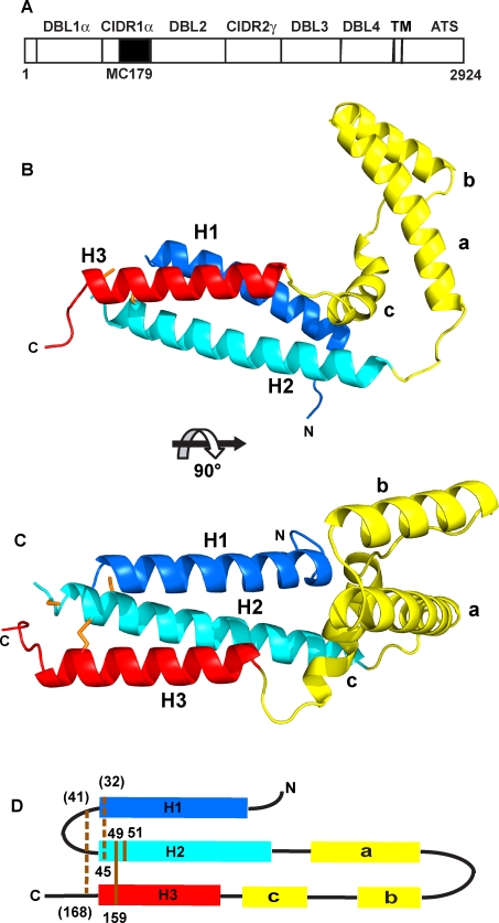
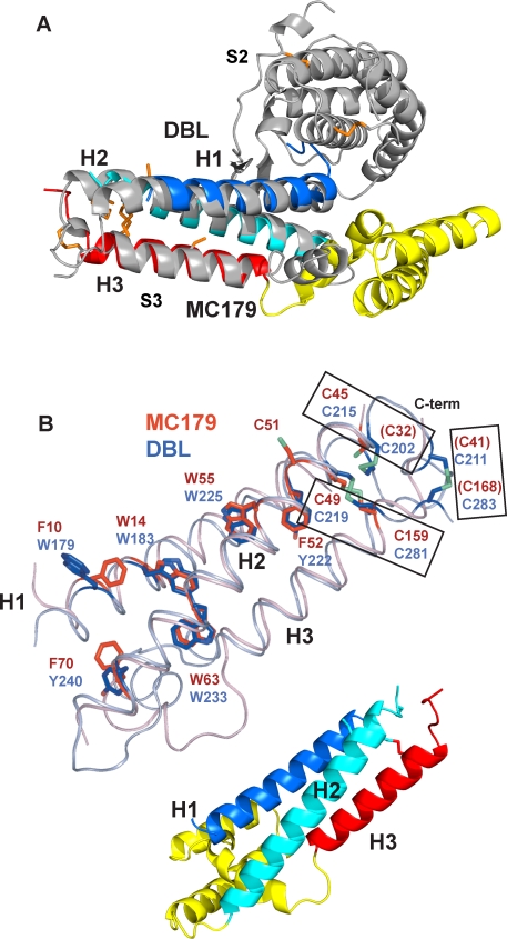
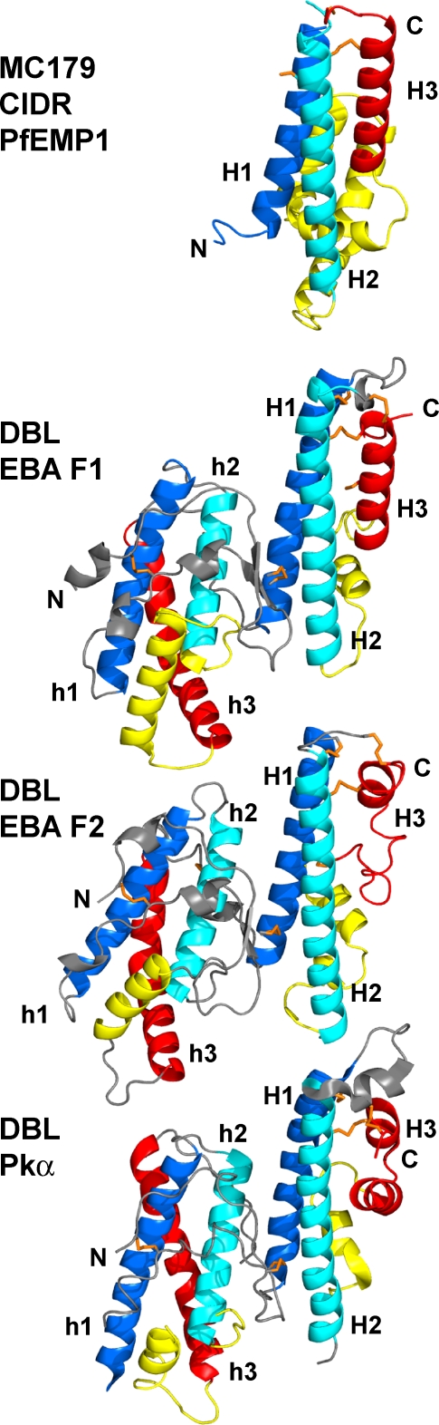
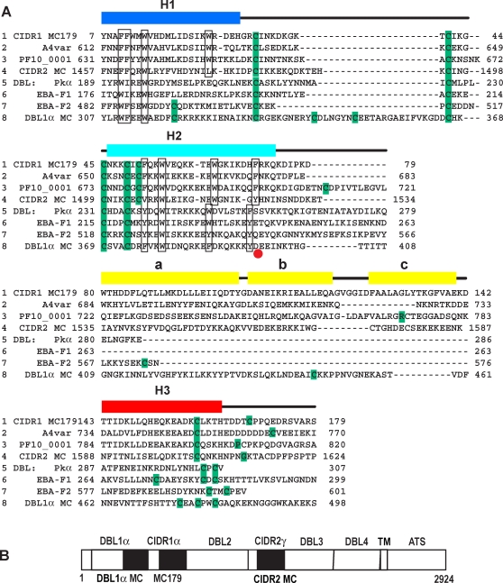
Plasmodium falciparum malaria parasites, living in red blood cells, express proteins of the erythrocyte membrane protein-1 (PfEMP1) family on the red blood cell surface. The binding of PfEMP1 molecules to human cell surface receptors mediates the adherence of infected red blood cells to human tissues. The sequences of the 60 PfEMP1 genes in each parasite genome vary greatly from parasite to parasite, yet the variant PfEMP1 proteins maintain receptor binding. Almost all parasites isolated directly from patients bind the human CD36 receptor. Of the several kinds of highly polymorphic cysteine-rich interdomain region (CIDR) domains classified by sequence, only the CIDR1alpha domains bind CD36. Here we describe the CD36-binding portion of a CIDR1alpha domain, MC179, as a bundle of three alpha-helices that are connected by a loop and three additional helices. The MC179 structure, containing seven conserved cysteines and 10 conserved hydrophobic residues, predicts similar structures for the hundreds of CIDR sequences from the many genome sequences now known. Comparison of MC179 with the CIDR domains in the genome of the P. falciparum 3D7 strain provides insights into CIDR domain structure. The CIDR1alpha three-helix bundle exhibits less than 20% sequence identity with the three-helix bundles of Duffy-binding like (DBL) domains, but the two kinds of bundles are almost identical. Despite the enormous diversity of PfEMP1 sequences, the CIDR1alpha and DBL protein structures, taken together, predict that a PfEMP1 molecule is a polymer of three-helix bundles elaborated by a variety of connecting helices and loops. From the structures also comes the insight that DBL1alpha domains are approximately 100 residues larger and that CIDR1alpha domains are approximately 100 residues smaller than sequence alignments predict. This new understanding of PfEMP1 structure will allow the use of better-defined PfEMP1 domains for functional studies, for the design of candidate vaccines, and for understanding the molecular basis of cytoadherence.


Conflict of interest statement

The authors have declared that no competing interests exist.

Figures

Figure 1
Figure 1. The MC179 structure and PfEMP1.
(A) Two CIDR and four DBL domains compose the extracellular portion of the 2924-amino acid residue PfEMP1 molecule from the Malayan Camp (MC) strain of P. falciparum that is expressed on the surface of an infected red blood cell. MC179 (black box) is about 65% of the MC CIDR1α domain. Like most PfEMP1 molecules, the MC PfEMP1 has a semi-conserved N-terminal DBL1α-CIDR1α pair or "head structure" . The transmembrane (TM) segment and acidic terminal segment (ATS) are located inside the infected red blood cell. (B) The MC179 structure is made up of three helices (H1, dark blue; H2, light blue; H3, red) that form a three-helix bundle and three additional helices (a, b, c; yellow) that connect H2 to H3. (C) View of MC179 after reorienting by 90°. Cysteine side chains are shown in orange. (D) MC179 topology diagram shows how the six helices are connected. Lines (orange) and residue numbers identify the positions of the three disulfide bonds and one unbonded cysteine. Two disulfide bonds were inferred to be present (dashed lines). Three cysteines (parentheses) were not observed in electron density. N- and C-termini (N, C) are labeled.
Figure 2
Figure 2. MC179 binding to CD36.
Refolded MC179 bound to CD36-transfected CHO cells as assayed by flow cytometry ([A,B]; blue hatched peaks). (A) Incubation of MC179 with CD36-Fc fusion protein (100 μg/ml) diminished MC179 binding to CD36 on cells (orange peak). MC179 binding was revealed with anti-MC179 Aotus immune serum. (B) MC179 binding to CD36 is inhibited by each of three anti-CD36 mAbs (peaks outlined in red, violet, and green). The violet peak is under the red peak and is only partially visible. Binding of MC179 was visualized with an anti-pentaHis mAb. In both panels, controls used untransfected cells (peaks outlined in black). In (A), Aotus preimmune serum yielded results (not shown) identical to the untransfected control. (C) Ribbon diagrams of MC179 in the same orientations as (D) and (E). Three-helix bundles are located towards the center of the figure and connecting helices are at the sides. (D) Two views of the hydrophobic surface 180° apart show a hydrophobic patch (green, at right) on the connecting helices of MC179. Labeled residues are those reported to affect CD36-binding (see text). (E) Surface views of MC179 showing the distribution of positive (blue) and negative (red) surface charge. The negatively charged region (red, at left) on the connecting helices is on the opposite face from the hydrophobic patch at the right in (D).
Figure 3
Figure 3. Sequence conservation of all CIDR domains of the 3D7 genome mapped on to the MC179 structure.
High to low conservation is mapped as a color gradient (dark blue to white) on the molecular surface of MC179. A highly conserved surface patch (dark blue) means that a high percentage of the residues at that position in the alignment of all 3D7 CIDR domains (Figure S2) are identical to the surface-exposed MC179 residue. (A) View of one side of MC179 reveals little conservation. (B) View after a rotation of 180° shows a highly conserved region on the molecule. Cys 51 is conserved in all CIDR domains. Other highly conserved residues that contribute to the conserved surface between Cys 51 and the N-terminus are Trp 14, Asp 21, Trp 55, Lys 59, Glu 62, and Ile 66 (not shown). Little conservation is seen on the surfaces of the connecting helices. The cartoons to the right are reference diagrams in the same orientations as the surfaces. A few highly conserved residues are not visible at the surface because they are completely buried. Sequence alignments (Figures S1, S2 and S3) were used as input to the Protskin server to produce this figure and Figure 4.
Figure 4
Figure 4. Comparison of CIDR1α and CIDR2β conserved surfaces.
Sequence conservation (Figures S1 and S3) has been mapped as in Figure 3. MC179 has been rotated about 90° from the orientation in Figure 3 to show the region between the H1 and H3 helices. At left, a bright red patch means that a high percentage of 3D7 CIDR1α domains contain the same residue at the particular position as does MC179. In the middle, a bright violet patch means that a high percentage of 3D7 CIDR2β domains contain the same residue at the particular position as does MC179. Appearing at the surface of both types of domain are Cys 45, Cys 49, and Cys 159 that form part of the conserved disulfide bond network and Leu 19 and Leu 148 that make a conserved interaction between H1 and H3. The unusual nearly buried Ser 22-Glu 152 pair is unique to CIDR1α domains. At position 147, a conserved lysine is found in CIDR1α domains, but is a cysteine in most CIDR2β domains. At right is a reference diagram for the orientation of MC179 in this figure.
Figure 5
Figure 5. CIDR and DBL domains have structurally identical three-helix bundles.
(A) The three-helix bundle of MC179 closely overlays the C-terminal three-helix bundle of the F1 DBL domain of EBA-175 (gray). MC179 superimposes similarly on the F2 DBL domain of EBA-175 and the Pkα-DBL domain (not shown). Note that the connecting helices (yellow) extend between H2 and H3, but the DBL subdomain–subdomain interaction is approximately 120° away between the H1 and H2 helices. This overlay models our prediction of the DBL1α C-terminal three-helix bundle, with the yellow connecting helices of MC179 modeling the predicted connecting helices of DBL1α (see text). DBL subdomain 2 (S2) and subdomain 3 (S3) are labeled . (B) The three-helix bundles of MC179 (pink helices) and of the F1 DBL domain (steel blue helices) are superimposed, after an approximate 180° reorientation from (A) about the vertical axis. The connecting helices between H2 and H3 are not shown for clarity. Note the almost identical positions of the helices and of conserved Phe, Trp, Tyr, and Cys side chains (red, MC179; blue, F1 DBL). Cysteines making conserved disulfide bonds (rectangles) are from MC179 (red font) and from the F1 DBL domain (blue font). Parentheses enclose the three cysteines not observed in the MC179 electron density. Cartoon diagram serves as a reference for the orientation of MC179 in (B).
Figure 6
Figure 6. The three-helix bundles of MC179 and the DBL domains.
All four structures were aligned using the alpha carbon atoms of the H1 and H2 helices, then were translated apart for viewing. In subdomain 3, the similarity of the H1 (dark blue) and H2 (light blue) helices is apparent, but the H3 (red) helices differ among molecules. MC179 and the EBA-175 F1 DBL each contain regular H3 helices, but the F2 and Pkα DBL domains have irregular H3 helices. Nevertheless, these degenerate helices meander to a pair of conserved cysteines that make conserved bonds to H1 and H2, as in MC179 and in the F1 domain. DBL domains contain another three-helix bundle (h1, h2, h3) in the N-terminal half (subdomain 2) of the molecule that is colored here analogously to the MC179 H1, H2, H3, and connecting helices. Several characteristics of these N-terminal bundles suggest their relatedness to the MC179 bundle and to the C-terminal DBL bundles (see text). N- and C-termini (N, C) are labeled.
Figure 7
Figure 7. Sequence comparison of MC179, CIDR, and DBL domains.
(A) Alignment of four CIDR and four DBL domains with the lines numbered 1–8 for clarity of discussion. Lines 1–4 show MC179 and the MC179-like portions of three other CIDR domains: line 1, CIDR1α MC179; line 2, CIDR1α of A4var PfEMP1; line 3, CIDR1α of PF10_0001 of 3D7; and line 4, CIDR2γ of the MC strain (see [B]). Lines 5–8 show four DBL domains: line 5, Pkα; line 6, EBA-F1; line 7, EBA-F2; and line 8, DBL1α of MC PfEMP1. The sequence alignment is based on conserved cysteines (green), conserved hydrophobic residues (boxed), and on superimposed X-ray structures. Note the differing lengths and high sequence diversity in regions of lines 2–8 that are aligned with the a, b, and c helices (yellow) that connect H2 and H3 of MC179. The short connection between the H2 and H3 helices in the non-PfEMP1 DBL domains (lines 5–7) contrasts with the much longer connection observed in the MC179 structure (line 1), predicted in other CIDR domains (lines 2–4), and predicted in DBL1α (line 8). Note the long loop in DBL1α that connects the H1 and H2 helices. The end of the H2 helix is marked (red dot). Helices from the MC179 structure are positioned over the MC179 sequence in line 1. MC179 is numbered 1–179 based on the PDB accession code 3C64, which is the same sequence as residues 576–754 of GenBank U27338. The other sequences are numbered as in their GenBank or PDB depositions with accession codes: A4var, L42244; PF10_0001; CIDR2 MC, U27338; Pkα, 2C6L; EBA-F1, 1ZRO; EBA-F2, 1ZRO; DBL1α MC, U27338. (B) Diagram of the entire PfEMP1 from the MC strain showing the alternating DBL and CIDR domains as in Figure 1. Filled boxes denote locations of the "DBL1α MC," "CIDR1 MC179," and "CIDR2 MC" sequences that are aligned in (A).
Figure 8
Figure 8. MC179 forms a dimer in the crystal.
(A) Contact between the molecules is primarily between the helices (yellow) that connect the H2 and H3 helices and between the H1 (dark blue) to H3 (red) sides of the three-helix bundles. (B) The molecular surface of the crystalline dimer shows the intertwining of the two molecules in a "handshake" manner. One molecule (at right) is colored by sequence conservation in a ramp of blue to white as in Figure 3, and the other molecule (at left) is colored gray for contrast. The total buried solvent-accessible surface area is 4300 Å2. The surface complementarity is 0.70 and 55 residues (220 atoms) from each molecule take part in the interaction. The two MC179 molecules are related by a crystallographic two-fold symmetry axis. The locations of several residues are indicated.

References

    1. Kyes S, Horrocks P, Newbold C. Antigenic variation at the infected red cell surface in malaria. Annu Rev Microbiol. 2001;55:673–707. - PubMed
    1. Yazdani SS, Mukherjee P, Chauhan VS, Chitnis CE. Immune responses to asexual blood-stages of malaria parasites. Curr Mol Med. 2006;6:187–203. - PubMed
    1. Kraemer SM, Smith JD. A family affair: var genes, PfEMP1 binding, and malaria disease. Curr Opin Microbiol. 2006;9:374–380. - PubMed
    1. Ralph SA, Scherf A. The epigenetic control of antigenic variation in Plasmodium falciparum. Curr Opin Microbiol. 2005;8:434–440. - PubMed
    1. Dzikowski R, Templeton TJ, Deitsch K. Variant antigen gene expression in malaria. Cell Microbiol. 2006;8:1371–1381. - PubMed

Publication types

Cite

AltStyle によって変換されたページ (->オリジナル) /