This site needs JavaScript to work properly. Please enable it to take advantage of the complete set of features!
Skip to main page content
U.S. flag

An official website of the United States government

Dot gov

The .gov means it’s official.
Federal government websites often end in .gov or .mil. Before sharing sensitive information, make sure you’re on a federal government site.

Https

The site is secure.
The https:// ensures that you are connecting to the official website and that any information you provide is encrypted and transmitted securely.

NIH NLM Logo
Log in
Access keys NCBI Homepage MyNCBI Homepage Main Content Main Navigation
. 2001 May:Chapter 19:Unit 19.1.
doi: 10.1002/0471142735.im1901s28.

Schistosomiasis

Affiliations

Schistosomiasis

F Lewis. Curr Protoc Immunol. 2001 May.

Abstract

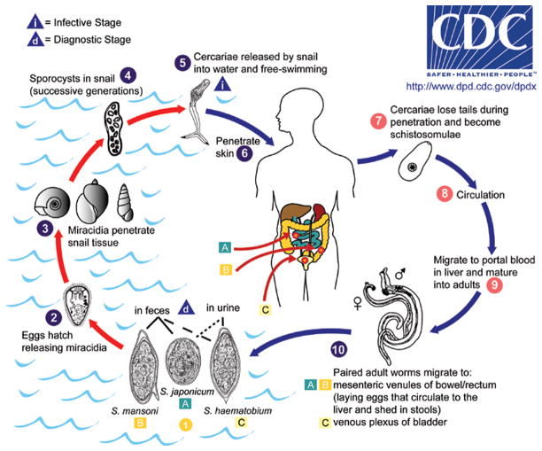
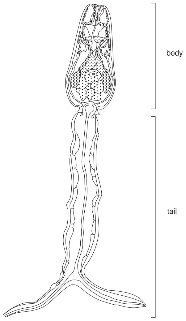
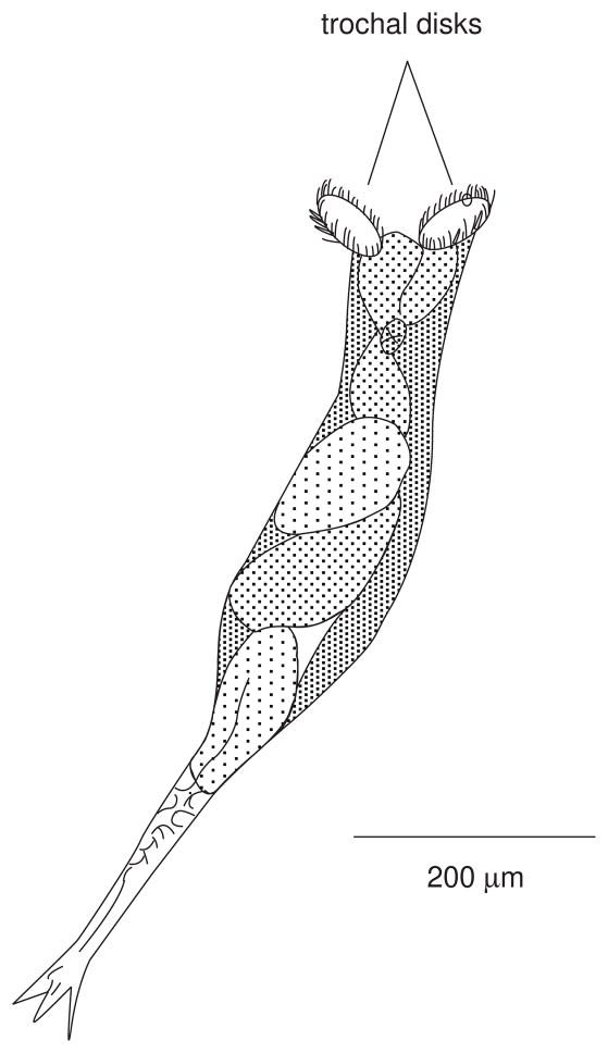
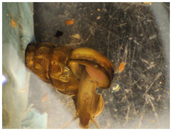
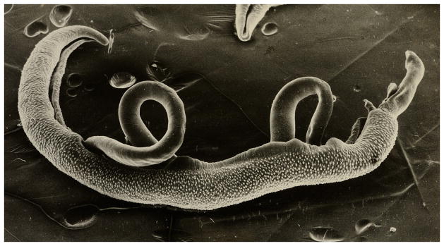
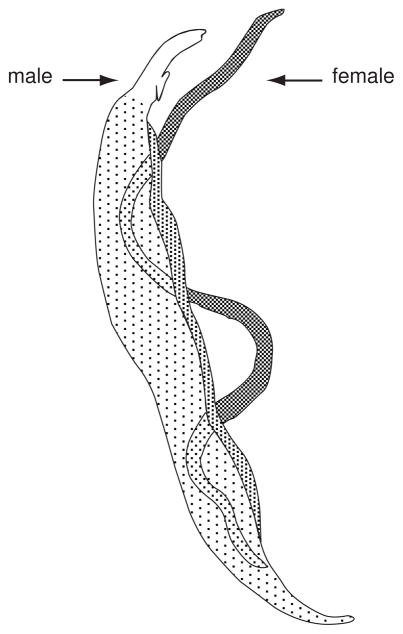
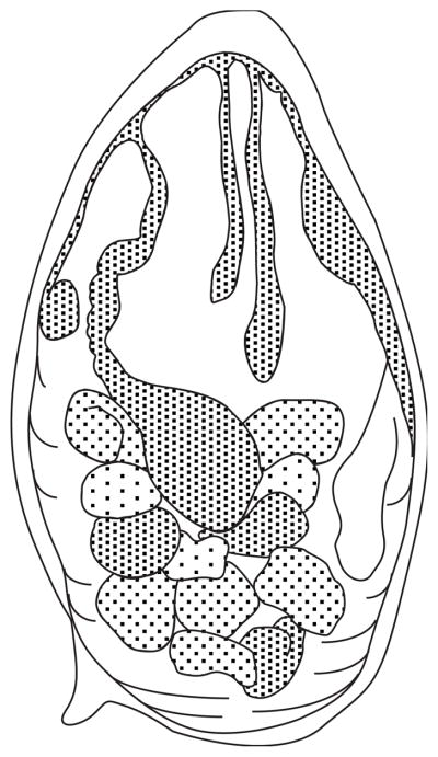
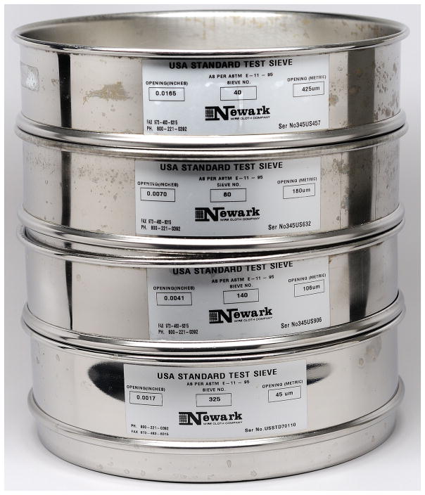
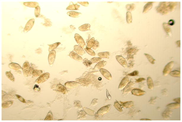
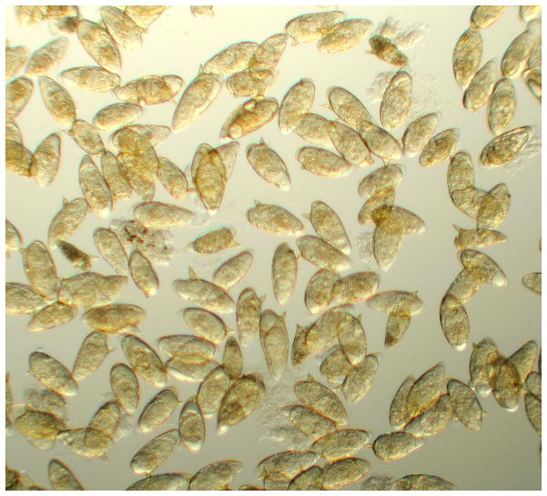
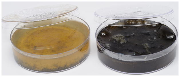
The trematode parasites in the family Schistosomatidae (phylum Platyhelminthes) infect a wide range of vertebrates. Three species of the genus Schistosoma are of major medical importance. This unit deals exclusively with the parasite Schistosoma mansoni, which is the species most frequently maintained in the laboratory. Among the far-ranging investigations in the immunology of schistosomiasis are studies in vaccine development, immunopathology of granulomatous inflammation and fibrosis, eosinophil function, and in vivo regulation of T(H)1 and T(H)2 responses. This unit describes maintenance and collection procedures for various stages of the schistosome that have immunologic interest, including infection of mice with cercariae, collection of cercariae, preparation of in vitro-derived schistosomules and in vivo-derived schistosomules, and collection of adult worms and eggs. Included also are techniques for preparing soluble egg antigen (SEA), one of the more commonly used schistosome antigenic preparations. A discussion is given of the basic steps that are important in maintaining the snail intermediate host, and infecting the snails with schistosome miracidia. The unit deals exclusively with the parasite Schistosoma mansoni, which is the species most frequently maintained in the laboratory. Since part of the life cycle of all schistosomes involves a snail host, a description of proper maintenance for the snails is provided. Often, problems in experiments can be traced back to improper snail and parasite maintenance, or lack of attention to detail during mammalian exposure to the infective stage (cercaria) of the parasite.


Figures

Figure 19.1.1
Figure 19.1.1
The life cycle of human schistosomes (image courtesy of the Centers for Disease Control and Prevention, DPDx). The figure depicts several life cycle stages that are mentioned in the text. For information on time to development of particular stages (e.g. lung schistosomules, adult worms), see the text for these specific stages. Key collection points mentioned in the text include: miracidia, cercariae, schistosomules, adult worms, and eggs. Approximate measurements for the various stages are: cercaria length (body plus tail), 500 μm; adult female worm length, 12 mm; adult male worm length, 9 mm; egg (length ×ばつ width), 140 ×ばつ 60 μm; miracidium (length ×ばつ width), 140 ×ばつ 55 μm.
Figure 19.1.2
Figure 19.1.2
Materials for mouse tail exposure to schistosome cercariae. Shown are a base and upper level of fluorescent light panels (available in standard hardware stores), separated by spacers and anchored with Velcro strips. The height is adjusted to 75 mm, to accommodate 75 ×ばつ 12 mm test tubes (not shown). A single mouse will be restrained in the Broome-type restrainer, with its tail extending from the bottom of the test tube that contains cercariae. Variations of these restrainers can be purchased commercially, or they can be engineered from plastics companies. Restrainers with diameter openings of about 25 mm can restrain mice up to 20 grams, whereas larger ones (up to 30 mm dia openings) are needed for larger mice.
Figure 19.1.3
Figure 19.1.3
Diagram of a Schistosoma mansoni cercaria. Since both body and tail are contractile, the overall length of this stage varies considerably, usually between 300 and 500 μm.
Figure 19.1.4
Figure 19.1.4
Components of the unit useful for filtering cercariae. The stainless steel screen has 47μm pore size openings, sufficient to allow passage of cercariae. The screen is placed inside of the lower filter portion and the clamp upper and lower parts together.
Figure 19.1.5
Figure 19.1.5
Sketch of a rotifer representative of the family Bdelloidea. The organisms are very contractile, and readily invert their trochal disks. When the disks are everted (as shown here) the movement of the cilia will resemble revolving wheels.
Figure 19.1.6
Figure 19.1.6
A male adult Oncomelania hupensis hupensis snail. The apex of the snail has been anchored in children’s clay, and the headfoot is extended. Extending from the right edge of the shell opening is a flesh-colored structure (the verge).
Figure 19.1.7
Figure 19.1.7
Typical collection of soil in which Oncomelania hupensis ssp. will lay their eggs. Magnified here approximately 150X, it is difficult to distinguish Oncomelania hupensis ssp. eggs from the surrounding granules, without agitating the dish and observing the less dense eggs moving more easily than the granules.
Figure 19.1.8
Figure 19.1.8
Three Oncomelania hupensis ssp. eggs. The eggs are covered in mud. Two of them were removed from the plastic surface of their pan, hence the flat surface of the egg can easily be observed.
Figure 19.1.9
Figure 19.1.9
1-liter side arm flask, painted black with the exception of approx 20 mm of the upper section of its side arm. Once eggs are deposited in the flask and it is filled with aged tap water, with a light source directed at the clear side arm, miracidia will concentrate in the clear side arm within 20–30 minutes. This allows easy collection, with a Pasteur pipette, of a relatively clean miracidial preparation
Figure 19.1.10
Figure 19.1.10
Electron micrograph of adult male and female Schistosoma mansoni in copula. The female is residing in the gynecophoral canal of the male worm. Length of males ranges from 6 to 13 mm; that of females ranges from 10 to 20 mm.
Figure 19.1.11
Figure 19.1.11
Sketch of perfusion apparatus for collecting adult Schistosoma mansoni from the mesenteric venous system. A 20-gauge needle, fitted onto the end of Tygon tubing, is placed in the descending aorta. Adult worms are collected in the perfusate from the severed portal vein.
Figure 19.1.12
Figure 19.1.12
Sketch of a Schistosoma mansoni egg. Approximate dimensions are 140 μm (length) by 60 μm (width).
Figure 19.1.13
Figure 19.1.13
Stainless steel sieves stacked (top to bottom) with pore sizes 420 μm, 180 μm, 105 μm, and 45 μm. Once the tissue homogenate is placed in the top-most sieve, repeated washing of the column with 1.2% NaCl results in eggs trapped on the surface of the lowest sieve, relatively free of contaminating larger particulate matter.
Figure 19.1.14
Figure 19.1.14
Falcon 2340 Cell Strainer. These commercially-available screens work well for concentrating schistosome eggs. Pore size of 40 μm allows retention of the eggs on the bottom of the screen, while extraneous, smaller matter passes through.
Figure 19.1.15
Figure 19.1.15
Sample of S. mansoni eggs collected from the surface of the wire cloth retaining sieve (45-μm pore size). The suspension contains considerable amounts of extraneous tissue at this stage in purification.
Figure 19.1.16
Figure 19.1.16
Sample of S. mansoni eggs after 3–4 cycles of swirling for concentration of mostly mature eggs.
Figure 19.1.17
Figure 19.1.17
Freshly seeded mud base plate with Nostoc sp. (left). On the right is an identical plate placed under a fluorescent light, at room temperature, for 3 weeks. Notice the dark mat of Nostoc sp. on the surface of the plate.

References

    1. Basch PF. Cultivation of Schistosoma mansoni in vitro. I Establishment of cultures from cercariae and development until pairing. J Parasitol. 1981;67:179–185. - PubMed
    1. Boros DL, Warren KS. Delayed hypersensitivity granuloma formation and dermal reaction induced and elicited by a soluble factor isolated from Schistosoma mansoni eggs. J Exp Med. 1970;132:488–507. - PMC - PubMed
    1. Bruce JI, Liang YS. Cultivation of schistosomes and snails for researchers in the United States of America and other countries. J Med Appl Malacol. 1992;4:13–30.
    1. Bruce JI, Radke MG, Davis GM. Biomedical Report No 19,406th Medical Laboratory. U.S. Army; 1971. Culturing Biomphalaria and Oncomelania (Gastropoda) for large-scale studies of schistosomiasis.
    1. Cai Y, Langley JG, Smith DI, Boros DL. A cloned major Schistosoma mansoni egg antigen with homologies to small heat shock proteins elicits Th1 responsiveness. Infect Immun. 1996;64:1750–1755. - PMC - PubMed

Substances

Cite

AltStyle によって変換されたページ (->オリジナル) /