This site needs JavaScript to work properly. Please enable it to take advantage of the complete set of features!
Skip to main page content
U.S. flag

An official website of the United States government

Dot gov

The .gov means it’s official.
Federal government websites often end in .gov or .mil. Before sharing sensitive information, make sure you’re on a federal government site.

Https

The site is secure.
The https:// ensures that you are connecting to the official website and that any information you provide is encrypted and transmitted securely.

NIH NLM Logo
Log in
Access keys NCBI Homepage MyNCBI Homepage Main Content Main Navigation
. 2008 Feb 12;105(6):1999-2004.
doi: 10.1073/pnas.0711799105. Epub 2008 Feb 1.

Adaptations of Trypanosoma brucei to gradual loss of kinetoplast DNA: Trypanosoma equiperdum and Trypanosoma evansi are petite mutants of T. brucei

Affiliations

Adaptations of Trypanosoma brucei to gradual loss of kinetoplast DNA: Trypanosoma equiperdum and Trypanosoma evansi are petite mutants of T. brucei

De-Hua Lai et al. Proc Natl Acad Sci U S A. .

Abstract

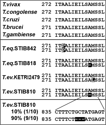
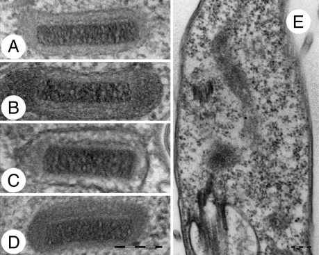
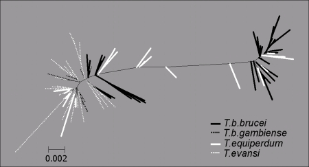
Trypanosoma brucei is a kinetoplastid flagellate, the agent of human sleeping sickness and ruminant nagana in Africa. Kinetoplastid flagellates contain their eponym kinetoplast DNA (kDNA), consisting of two types of interlocked circular DNA molecules: scores of maxicircles and thousands of minicircles. Maxicircles have typical mitochondrial genes, most of which are translatable only after RNA editing. Minicircles encode guide RNAs, required for decrypting the maxicircle transcripts. The life cycle of T. brucei involves a bloodstream stage (BS) in vertebrates and a procyclic stage (PS) in the tsetse fly vector. Partial [dyskinetoplastidy (Dk)] or total [akinetoplastidy (Ak)] loss of kDNA locks the trypanosome in the BS form. Transmission between vertebrates becomes mechanical without PS and tsetse mediation, allowing the parasite to spread outside the African tsetse belt. Trypanosoma equiperdum and Trypanosoma evansi are agents of dourine and surra, diseases of horses, camels, and water buffaloes. We have characterized representative strains of T. equiperdum and T. evansi by numerous molecular and classical parasitological approaches. We show that both species are actually strains of T. brucei, which lost part (Dk) or all (Ak) of their kDNA. These trypanosomes are not monophyletic clades and do not qualify for species status. They should be considered two subspecies, respectively T. brucei equiperdum and T. brucei evansi, which spontaneously arose recently. Dk/Ak trypanosomes may potentially emerge repeatedly from T. brucei.


Conflict of interest statement

The authors declare no conflict of interest.

Figures

Fig. 1.
Fig. 1.
Parasitaemia growth curves. (A) Growth curve of parasitaemia caused by representative strains in mice. The cell density in blood is indicated. (B) Growth curve of parasitaemia of the same strains as in A in mice with concurrent i.p. injection of 10 μg of EtdBr per gram of weight.
Fig. 2.
Fig. 2.
Minicircle homogenization (A) and/or absence (B). (A) EtdBr-stained agarose gel showing restriction analysis of kDNA minicircles from representative strains using MboI (see also SI Fig. 11). Linearized minicircles are indicated by an arrowhead. Note the presence of kDNA network in the slot of the T. brucei lane (arrow). (B) Southern blotting of total DNA isolated from representative strains and digested with restriction enzyme TaqI. The membrane was hybridized with a radiolabeled probe prepared from total T. brucei minicircles.
Fig. 3.
Fig. 3.
Transcript levels (A) and RNA editing (B). (A) Levels of mt and cytosolic transcripts detected by Northern blotting in representative strains of T. brucei, T. equiperdum, and T. evansi. P, preedited; E, edited; A6, ATP synthase subunit 6; cox1–3, cytochrome oxidase subunits 1–3; trCO4, trypanosome cytochrome oxidase subunit 4; CO6, cytochrome oxidase subunit 6; 18S, 18S ribosomal RNA. As a control, the gel was stained with EtdBr to visualize rRNA bands. (B) RNA editing of some mRNAs is affected in the Dk/Ak cells. Real-time PCR analysis of preedited, edited, and never-edited mRNAs, performed in triplicate on cDNAs. For each target amplicon, the relative change in RNA abundance was determined by using cytosolic transcripts of β-tubulin and 18S rRNA (data not shown) as internal references, because their transcription was not affected. The relative abundance of each examined transcript was plotted on a logarithmic scale: 1.0 represents the level in BS of T. brucei STIB920; A6, ATPase subunit 6; cox2, cytochrome oxidase subunit 2; MURF2, maxicircle unknown reading frame 2; RPS12, ribosomal protein S12; never-edited cox1 mRNA; and ND4, NADH-dehydrogenase subunit 4.
Fig. 4.
Fig. 4.
Protein levels (A) and respiration (B). (A) Levels of mt and cytosolic proteins detected by Western blotting in strains of T. brucei, T. equiperdum, and T. evansi. MRP1/2, mtRNA-binding proteins 1/2; TbRGG1, editing-associated RGG1 protein; RBP16, RNA-binding protein 16; TbTUT, terminal uridyl transferase; UMSBP, universal minicircle sequence binding protein; POL IC, mt DNA polymerase IC; apo C1, apocytochrome Cyt C1; Cyt C, cytochrome c; Rieske, Rieske Fe-S protein; trCO4, trypanosome cytochrome oxidase subunit 4; CO6, cytochrome oxidase subunit 6; p18, ATP synthase subunit b; ATPase β, ATP synthase subunit β; hsp60, heat shock protein 60. (B) Relative contribution of TAO-mediated pathway (AP) and cytochrome-mediated pathway (CP) in T. brucei PS and BS, and the Dk/Ak strains. The amount of O2 consumption inhibited by KCN was measured as the CP capacity. The mean and the SD values of three experiments are shown.
Fig. 5.
Fig. 5.
Amino acid (Top and Middle) and nucleotide (Bottom) alignment of a highly conserved part of the γ subunit of ATP synthase.
Fig. 6.
Fig. 6.
Electron microscopy of kinetoplasts in fixed cells. (A) T. brucei STIB920, (B) T. equiperdum STIB842, (C) T. equiperdum STIB818, (D) T. evansi STIB810, and (E) T. evansi STIB805. (Scale bar, 200 nm.)
Fig. 7.
Fig. 7.
Neighbor-joining clustering of the SL RNA repeats from Trypanosoma species. Alignment was performed by using CLUSTALW with gap opening weight = 15 and gap extension weight = 6.6.

References

    1. Simpson AGB, Stevens JR, Lukeš J. The evolution and diversity of kinetoplastid flagellates. Trends Parasitol. 2006;22:168–174. - PubMed
    1. Lukeš J, Hashimi H, Zíková A. Unexplained complexity of the mitochondrial genome and transcriptome in kinetoplastid flagellates. Curr Genet. 2005;48:277–299. - PubMed
    1. Stuart KD, Schnaufer A, Ernst NL, Panigrahi AK. Complex management: RNA editing in trypanosomes. Trends Biochem Sci. 2005;30:97–105. - PubMed
    1. Shlomai J. The structure and replication of kinetoplast DNA. Curr Mol Med. 2004;4:623–647. - PubMed
    1. Liu BY, Liu YN, Motyka SA, Agbo EEC, Englund PT. Fellowship of the rings: the replication of kinetoplast DNA. Trends Parasitol. 2005;21:363–369. - PubMed

Publication types

Cite

AltStyle によって変換されたページ (->オリジナル) /