This site needs JavaScript to work properly. Please enable it to take advantage of the complete set of features!
Skip to main page content
U.S. flag

An official website of the United States government

Dot gov

The .gov means it’s official.
Federal government websites often end in .gov or .mil. Before sharing sensitive information, make sure you’re on a federal government site.

Https

The site is secure.
The https:// ensures that you are connecting to the official website and that any information you provide is encrypted and transmitted securely.

NIH NLM Logo
Log in
Access keys NCBI Homepage MyNCBI Homepage Main Content Main Navigation
. 2008 Mar;294(3):C810-9.
doi: 10.1152/ajpcell.00511.2007. Epub 2008 Jan 23.

Apical maxi-K (KCa1.1) channels mediate K+ secretion by the mouse submandibular exocrine gland

Affiliations

Apical maxi-K (KCa1.1) channels mediate K+ secretion by the mouse submandibular exocrine gland

Tetsuji Nakamoto et al. Am J Physiol Cell Physiol. 2008 Mar.

Abstract

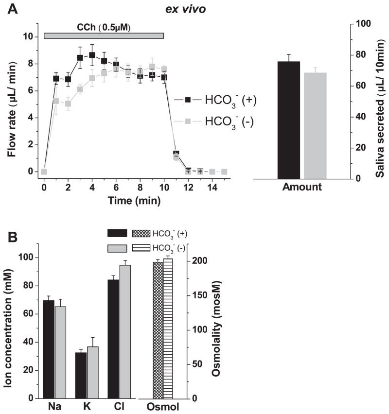
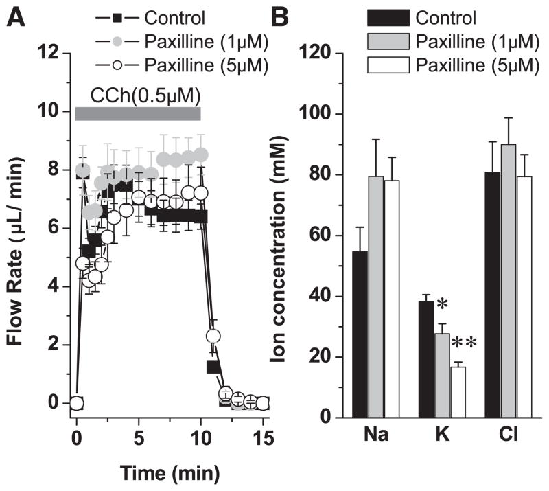
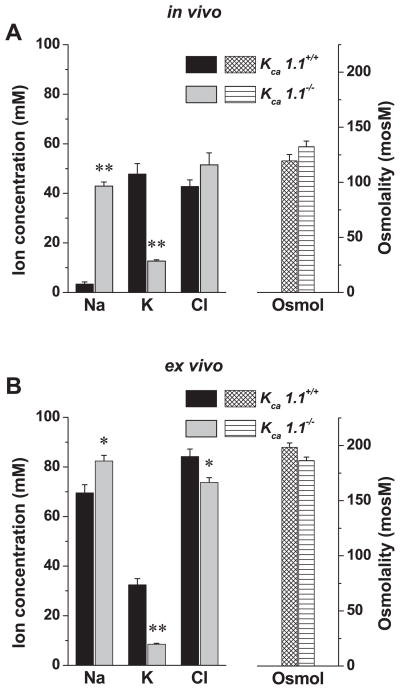
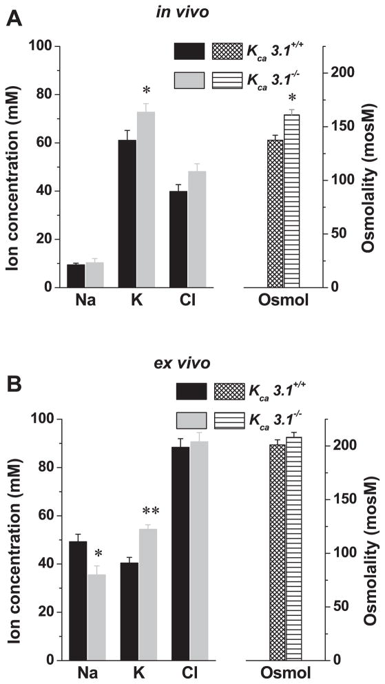
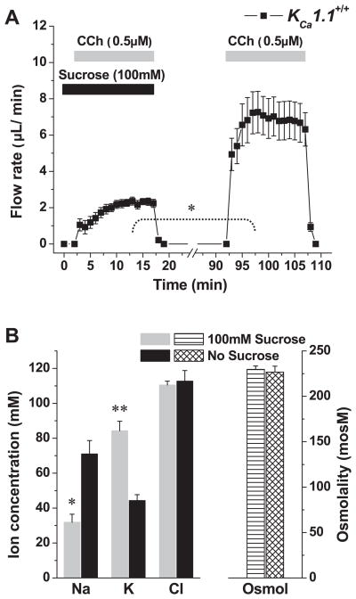
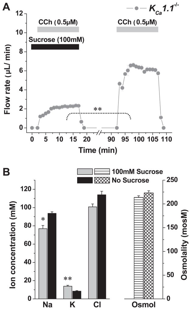
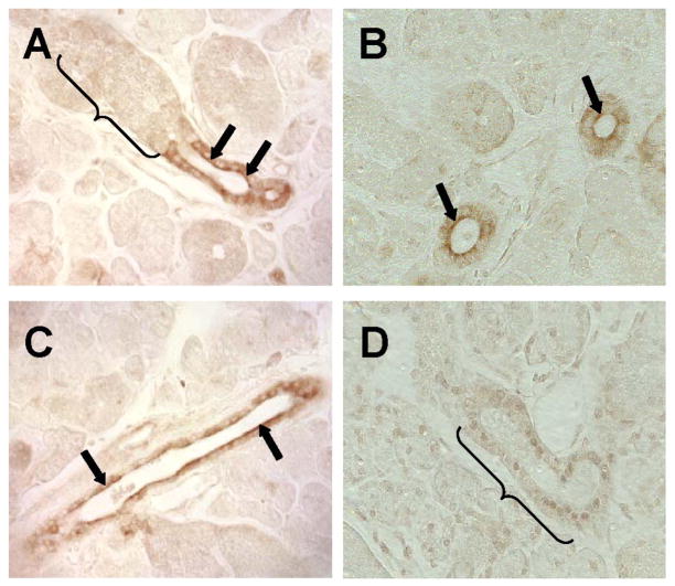
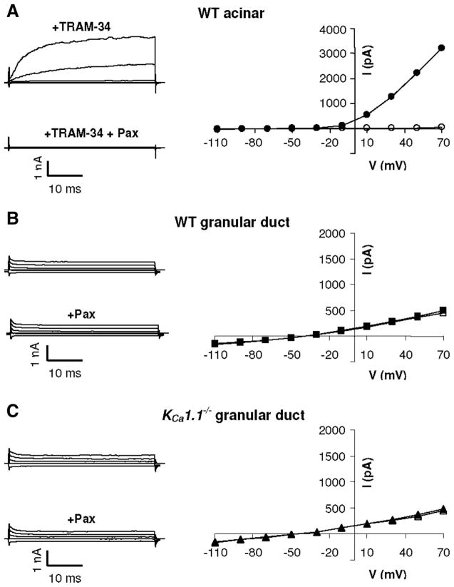
The exocrine salivary glands of mammals secrete K+ by an unknown pathway that has been associated with HCO3(-) efflux. However, the present studies found that K+ secretion in the mouse submandibular gland did not require HCO3(-), demonstrating that neither K+/HCO3(-) cotransport nor K+/H+ exchange mechanisms were involved. Because HCO3(-) did not appear to participate in this process, we tested whether a K channel is required. Indeed, K+ secretion was inhibited >75% in mice with a null mutation in the maxi-K, Ca2+-activated K channel (KCa1.1) but was unchanged in mice lacking the intermediate-conductance IKCa1 channel (KCa3.1). Moreover, paxilline, a specific maxi-K channel blocker, dramatically reduced the K+ concentration in submandibular saliva. The K+ concentration of saliva is well known to be flow rate dependent, the K+ concentration increasing as the flow decreases. The flow rate dependence of K+ secretion was nearly eliminated in KCa1.1 null mice, suggesting an important role for KCa1.1 channels in this process as well. Importantly, a maxi-K-like current had not been previously detected in duct cells, the theoretical site of K+ secretion, but we found that KCa1.1 channels localized to the apical membranes of both striated and excretory duct cells, but not granular duct cells, using immunohistochemistry. Consistent with this latter observation, maxi-K currents were not detected in granular duct cells. Taken together, these results demonstrate that the secretion of K+ requires and is likely mediated by KCa1.1 potassium channels localized to the apical membranes of striated and excretory duct cells in the mouse submandibular exocrine gland.


Figures

Fig. 1
Fig. 1
Effects of HCO3 depletion on fluid secretion and ion composition in ex vivo submandibular glands of wild-type KCa1.1 mice. Isolated, perfused submandibular glands (see MATERIALS AND METHODS) were used to determine the effects of HCO3 depletion on the ion composition of saliva. Carbachol (CCh, 0.5 μM) was used to stimulate secretion. Flow rate was expressed as μl/min. HCO3-free solutions contained 100 μM of the carbonic anhydrase inhibitor acetazolamide. A: saliva flow rate (left) and total amount saliva secreted by 10-min stimulation (right). Glands were perfused with (black symbols, n = 10) and without (gray symbols, n = 4) external HCO3. B: ion composition and osmolality of the saliva shown in A. No significant difference in the ion concentration or osmolality was found between HCO3-containing and HCO3-free samples. Values represent means ± SE. KCa1.1, calcium-activated K channel isoform 1.1.
Fig. 2
Fig. 2
The effect of paxilline on ex vivo saliva flow rate and ion composition. A: ex vivo submandibular glands were perfused with paxilline (1 or 5 μM) for 30 min before stimulation with carbachol (0.5 μM) in the continued presence of paxilline. B: the ion composition was analyzed. The numbers of glands were control = 10, 1 μM paxilline = 4, and 5 μM paxilline = 6 from equal numbers of male and female wild-type mice; values represent means ± SE; *P < 0.05 and **P < 0.001. Wild-type mice used in this experiment were BlackSwiss-129 SvJ hybrid mice.
Fig. 3
Fig. 3
In vivo and ex vivo ion composition of submandibular saliva secreted by KCa1.1+/+ and KCa1.1−/− mice. A: in vivo salivation was induced by the muscarinic agonist pilocarpine (10 mg/kg, intraperitoneal injection), and the ion composition (left) and osmolality (right) of saliva collected over 30 min were analyzed (KCa1.1+/+, n = 12; KCa1.1−/−, n = 12). B: ex vivo salivation was induced by the muscarinic agonist carbachol (0.5 μM), and the ion composition (left) and osmolality (right) of saliva collected over 10 min were analyzed (KCa1.1+/+, n = 10; KCa1.1−/−, n = 5). Values represent means ± SE; *P < 0.05 and **P < 0.001.
Fig. 4
Fig. 4
In vivo and ex vivo ion composition of submandibular saliva secreted by KCa3.1+/+ and KCa3.1−/− mice. A: in vivo salivation was induced by the muscarinic agonist pilocarpine (10 mg/kg, intraperitoneal injection), and the ion composition (left) and osmolality (right) of saliva collected over 30 min were analyzed (KCa3.1+/+, n = 20; KCa3.1−/−, n = 22). B: ex vivo salivation was induced by the muscarinic agonist carbachol (0.5 μM), and the ion composition (left) and osmolality (right) of saliva collected over 10 min were analyzed (KCa3.1+/+, n = 20; KCa3.1−/−, n = 20). Values represent means ± SE; *P < 0.05 and **P < 0.001.
Fig. 5
Fig. 5
Effects of hyperosmotic challenge on saliva flow and ion composition in the ex vivo submandibular glands of wild-type KCa1.1 mice. A: glands were stimulated with the muscarinic agonist carbachol (0.5 μM) in the presence or absence of 100 mM sucrose as indicated (n = 5). B: ion concentrations and osmolality of the saliva collected in the experiments shown in A. Values represent means ± SE; *P < 0.05 and **P < 0.001.
Fig. 6
Fig. 6
Effects of hyperosmotic challenge on saliva flow and ion composition in the ex vivo submandibular glands of wild-type KCa1.1−/− mice. A: glands were stimulated with the muscarinic agonist carbachol (0.5 μM) in the presence or absence of 100 mM sucrose as indicated (n = 6). B: ion concentrations and osmolality of the saliva collected in the experiments shown in A. Values represent means ± SE; *P < 0.05 and **P < 0.001. Note that ~90% of the hypertonic-induced K+ secretion was inhibited in KCa1.1−/− mice (compare with Fig. 5).
Fig. 7
Fig. 7
Localization of the KCa1.1 channel in mouse submandibular glands. Immunohistochemical staining of the KCa1.1 channel was observed in the apical region of striated (arrows in A and B) and excretory (arrows in C) ducts, but not in granular duct cells in wild-type KCa1.1 mice (bracket in A). D: no staining was detectable in the striated duct cells of KCa1.1−/− mice (bracket).
Fig. 8
Fig. 8
Cationic current in mouse submandibular gland cells. Single acinar (A) or granular duct (B and C) cells were isolated from submandibular glands of either wild-type (WT; A and B) or KCa1.1-null (C) mice. Cationic currents were measured in the presence of 250 nM intracellular Ca2+ (solid symbols) and then treated with 1 μM of the maxi-K specific inhibitor paxilline (Pax; open symbols). The acinar cell currents in A were recorded in the presence of the KCa3.1-specific inhibitor TRAM-34 (1 μM). The solid and open symbols in B and C mostly overlie each other. V, voltage; I, current.

References

    1. Alexander JH, van Lennep EW, Young JA. Water and electrolyte secretion by the exorbital lacrimal gland of the rat studied by micropuncture and catheterization techniques. Pflügers Arch. 1972;337:299–309. - PubMed
    1. Begenisich T, Nakamoto T, Ovitt CE, Nehrke K, Brugnara C, Alper SL, Melvin JE. Physiological roles of the intermediate conductance, Ca2+-activated potassium channel Kcnn4. J Biol Chem. 2004;279:47681–47687. - PubMed
    1. Botelho SY, Martinez EV. Electrolytes in lacrimal gland fluid and in tears at various flow rates in the rabbit. Am J Physiol. 1973;225:606–609. - PubMed
    1. Chaturapanich G, Ishibashi H, Dinudom A, Young JA, Cook DI. H+ transporters in the main excretory duct of the mouse mandibular salivary gland. J Physiol. 1997;503:583–598. - PMC - PubMed
    1. Cook DI, Van Lennep EW, Roberts ML, Young JA. Secretion by the major salivary glands. In: Johnson LR, editor. Physiology of the Gastrointestinal Tract. 3. New York: Raven Press; 1994. pp. 1061–1117.

Publication types

MeSH terms

Cite

AltStyle によって変換されたページ (->オリジナル) /