Crystal structure of activin receptor type IIB kinase domain from human at 2.0 Angstrom resolution
- PMID: 17893364
- PMCID: PMC2204126
- DOI: 10.1110/ps.073068407
Crystal structure of activin receptor type IIB kinase domain from human at 2.0 Angstrom resolution
Abstract
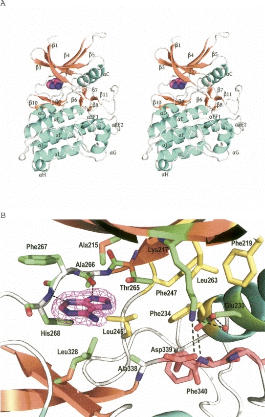
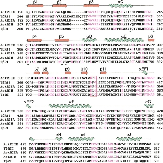
Activin receptor type IIB (ActRIIB), a type II TGF-beta serine/threonine kinase receptor, is integral to the activin and myostatin signaling pathway. Ligands such as activin and myostatin bind to activin type II receptors (ActRIIA, ActRIIB), and the GS domains of type I receptors are phosphorylated by type II receptors. Myostatin, a negative regulator of skeletal muscle growth, is regarded as a potential therapeutic target and binds to ActRIIB effectively, and to a lesser extent, to ActRIIA. The high-resolution structure of human ActRIIB kinase domain in complex with adenine establishes the conserved bilobal architecture consistent with all other catalytic kinase domains. The crystal structure reveals that the adenine has a considerably different orientation from that of the adenine moiety of ATP observed in other kinase structures due to the lack of an interaction by ribose-phosphate moiety and the presence of tautomers with two different protonation states at the N9 nitrogen. Although the Lys217-Glu230 salt bridge is absent, the unphosphorylated activation loop of ActRIIB adopts a conformation similar to that of the fully active form. Unlike the type I TGF-beta receptor, where a partially conserved Ser280 is a gatekeeper residue, the AcRIIB structure possesses Thr265 with a back pocket supported by Phe247. Taken together, these structural features provide a molecular basis for understanding the coupled activity and recognition specificity for human ActRIIB kinase domain and for the rational design of selective inhibitors.
Figures
References
-
- DeLano W.L. 2002. The PyMOL molecular graphics system, DeLano Scientific, San Carlos, CA, http://www.pymol.org.
-
- Donaldson C.J., Mathews, L.S., and Vale, W.W. 1992. Molecular cloning and binding properties of the human type II activin receptor. Biochem. Biophys. Res. Commun. 184: 310–316. - PubMed
-
- Graham H. and Peng, C. 2006. Activin receptor-like kinases: Structure, function and clinical implications. Endocr. Metab. Immune Disord. Drug Targets 6: 45–58. - PubMed
-
- Greenwald J., Vega, M.E., Allendorph, G.P., Fischer, W., and Choe, S. 2004. A flexible activin explains the membrane-dependent cooperative assembly of TGF-β family receptors. Mol. Cell 15: 485–489. - PubMed
MeSH terms
Substances
Associated data
- Actions
LinkOut - more resources
Full Text Sources