A previously unknown reovirus of bat origin is associated with an acute respiratory disease in humans
- PMID: 17592121
- PMCID: PMC1899191
- DOI: 10.1073/pnas.0701372104
A previously unknown reovirus of bat origin is associated with an acute respiratory disease in humans
Abstract
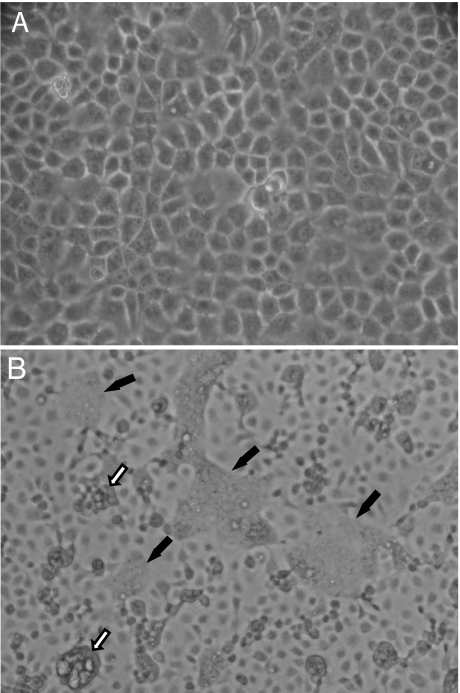
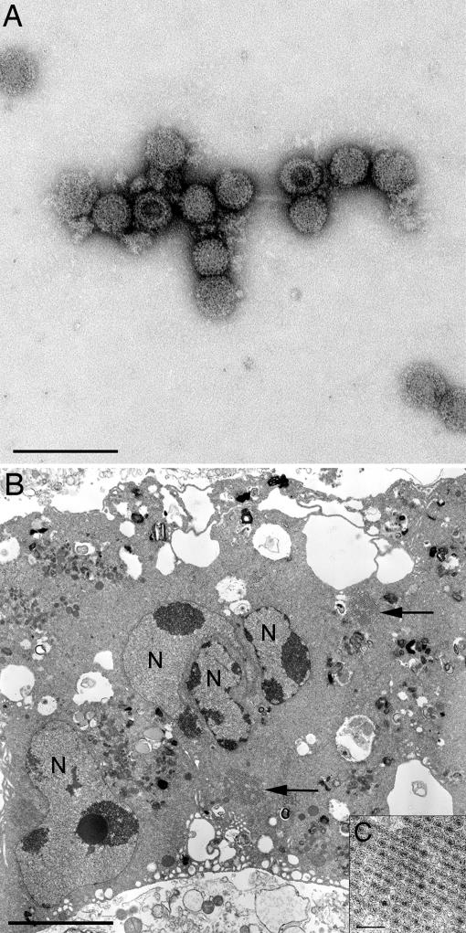
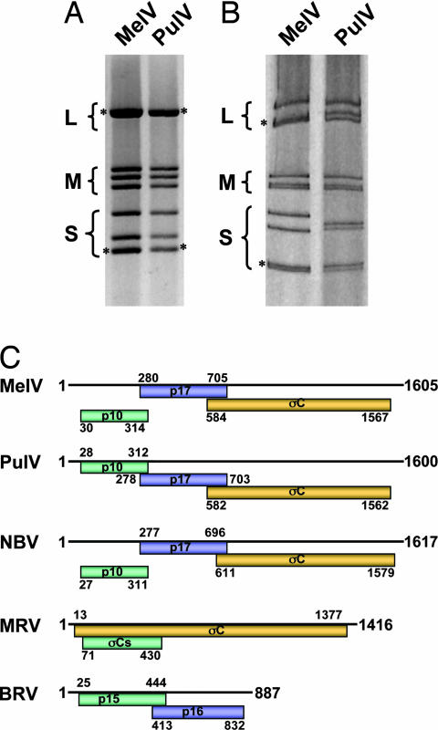
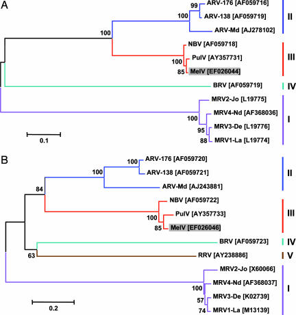
Respiratory infections constitute the most widespread human infectious disease, and a substantial proportion of them are caused by unknown etiological agents. Reoviruses (respiratory enteric orphan viruses) were first isolated from humans in the early 1950s and so named because they were not associated with any known disease. Here, we report a previously unknown reovirus (named "Melaka virus") isolated from a 39-year-old male patient in Melaka, Malaysia, who was suffering from high fever and acute respiratory disease at the time of virus isolation. Two of his family members developed similar symptoms approximately 1 week later and had serological evidence of infection with the same virus. Epidemiological tracing revealed that the family was exposed to a bat in the house approximately 1 week before the onset of the father's clinical symptoms. Genome sequence analysis indicated a close genetic relationship between Melaka virus and Pulau virus, a reovirus isolated in 1999 from fruit bats in Tioman Island, Malaysia. Screening of sera collected from human volunteers on the island revealed that 14 of 109 (13%) were positive for both Pulau and Melaka viruses. This is the first report of an orthoreovirus in association with acute human respiratory diseases. Melaka virus is serologically not related to the different types of mammalian reoviruses that were known to infect humans asymptomatically. These data indicate that bat-borne reoviruses can be transmitted to and cause clinical diseases in humans.
Conflict of interest statement
The authors declare no conflict of interest.
Figures
Comment in
-
Absence of Melaka-virus in European children with respiratory disease.Schildgen V, Rüngeler E, Tillmann R, Schildgen O. Schildgen V, et al. J Clin Virol. 2008 Jul;42(3):295-6. doi: 10.1016/j.jcv.200802003. Epub 2008 Mar 24. J Clin Virol. 2008. PMID: 18359270 Free PMC article. No abstract available.
References
-
- Nibert ML, Schiff LA. In: Fields Virology. Knipe DM, Howley PM, Griffin DE, Lamb RA, Martin MA, Roizman B, Straus SE, editors. Philadelphia: Lippincott Williams & Wilkins; 2001. pp. 1679–1728.
-
- Mertens PPC, Duncan R, Attoui H, Dermody TS. In: Virus Taxonomy: Eighth Report of the International Committee on Taxonomy of Viruses. Fauquet CM, Mayo MA, Maniloff J, Desselberger U, Ball LA, editors. San Diego: Elsevier Academic; 2005. pp. 447–454.
MeSH terms
Associated data
- Actions
- Actions
- Actions
- Actions
LinkOut - more resources
Full Text Sources
Other Literature Sources
Medical