This site needs JavaScript to work properly. Please enable it to take advantage of the complete set of features!
Skip to main page content
U.S. flag

An official website of the United States government

Dot gov

The .gov means it’s official.
Federal government websites often end in .gov or .mil. Before sharing sensitive information, make sure you’re on a federal government site.

Https

The site is secure.
The https:// ensures that you are connecting to the official website and that any information you provide is encrypted and transmitted securely.

NIH NLM Logo
Log in
Access keys NCBI Homepage MyNCBI Homepage Main Content Main Navigation
. 2007 Jun 19;104(25):10536-41.
doi: 10.1073/pnas.0703535104. Epub 2007 Jun 6.

Molecular sabotage of plant defense by aphid saliva

Affiliations

Molecular sabotage of plant defense by aphid saliva

Torsten Will et al. Proc Natl Acad Sci U S A. .

Abstract

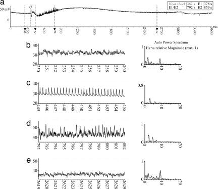
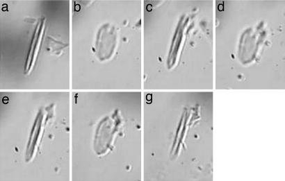
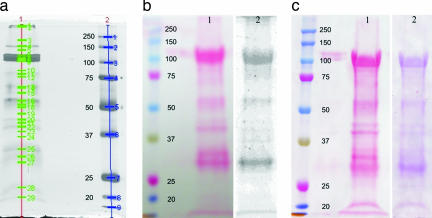
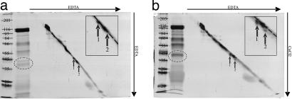
Aphids, which constitute one of the most important groups of agricultural pests, ingest nutrients from sieve tubes, the photoassimilate transport conduits in plants. Aphids are able to successfully puncture sieve tubes with their piercing mouthparts (stylets) and ingest phloem sap without eliciting the sieve tubes' normal occlusion response to injury. Occlusion mechanisms are calcium-triggered and may be prevented by chemical constituents in aphid saliva injected into sieve tubes before and during feeding. We recorded aphid feeding behavior with the electrical penetration graph (EPG) technique and then experimentally induced sieve tube plugging. Initiation of sieve tube occlusion caused a change in aphid behavior from phloem sap ingestion to secretion of watery saliva. Direct proof of "unplugging" properties of aphid saliva was provided by the effect of aphid saliva on forisomes. Forisomes are proteinaceous inclusions in sieve tubes of legumes that show calcium-regulated changes in conformation between a contracted state (below calcium threshold) that does not occlude the sieve tubes and a dispersed state (above calcium threshold) that occludes the sieve tubes. We demonstrated in vitro that aphid saliva induces dispersed forisomes to revert back to the nonplugging contracted state. Labeling Western-blotted saliva proteins with 45Ca2+ or ruthenium red inferred the presence of calcium-binding domains. These results demonstrate that aphid saliva has the ability to prevent sieve tube plugging by molecular interactions between salivary proteins and calcium. This provides aphids with access to a continuous flow of phloem sap and is a critical adaptation instrumental in the evolutionary success of aphids.


Conflict of interest statement

The authors declare no conflict of interest.

Figures

Fig. 1.
Fig. 1.
EPG recordings from sieve tube-related activities of the aphid M. viciae before, during, and after leaf heat shock in V. faba. (a) An overview trace of 1 hour, showing changes in the aphid's activities. (b–e) Four details of 10-sec duration at four different time intervals (time axis arrows b, c, d, and e in a). Auto power spectra present the waveform frequency (Hz) vs. the relative magnitude with a maximum of 1, which provide a major characteristic of waveform identity. Waveform E2 (b) represents ingestion of phloem sap accompanied by rhythmic secretion of small amounts of watery saliva that is ingested together with SE sap through the food canal; the watery saliva secreted during E2 is sucked up along with the sap and is believed to prevent clogging of the proteinaceous food components in the food canal (16, 23, 27, 28). Approximately 16 sec after heat shock is applied to the leaf tip (H in a) 6 cm from the feeding site, waveform E2 changes abruptly to E1 waveform, representing watery salivation into the SE lumen (20, 29) (c). Approximately 414 sec later, waveform E1 changes into a diffuse waveform (d), which may represent an irregular mixture of E1/E2 waveforms. Approximately 8.3 min after the heat shock, the aphid resumed its original feeding behavior (E2; e).
Fig. 2.
Fig. 2.
Reaction of an isolated forisome from V. faba sieve-tube to Ca2+, EDTA (a Ca2+ chelator), and watery saliva concentrate. (a) An isolated forisome in 10 μl of 2 mM EDTA extraction medium (25). (b) After application of 4 μl of 1 mM CaCl2, the forisome disperses and contracts again (c) after supplying 2 μl of 2 mM EDTA. (d and e) Repetition of this treatment has the same effect. (f) After the third application of 2 μl of 1 mM Ca2+, the forisome disperses again. (g) Subsequent application of 4 μl of saliva concentrate from M. viciae (from 6,750 aphids) leads to a contraction of the forisome comparable to that in response to addition of EDTA (c and e).
Fig. 3.
Fig. 3.
SDS/PAGE of the watery saliva of M. viciae and labeling of calcium-binding proteins with radioactive 45Ca2+ or by staining with ruthenium red on a Western blot nitrocellulose membrane. (a) Saliva concentrate of 1,000 aphids separated on a 10% gel (lane 1). Lane 2 presents marker proteins (kDa; Precision Plus Protein All Blue Standards; Bio-Rad, Hercules, CA). The silver-stained protein bands are marked and numbered by use of Quantity One 1-D Analysis Software (Bio-Rad). (b) Saliva collected from 41,000 aphids and separated on 10% SDS/PAGE and blotted on a nitrocellulose membrane. Lane 1 shows an overview of the separated proteins stained with Ponceau S, whereas the left lane presents marker proteins. In lane 2, after destaining the membrane, calcium-binding proteins are labeled with radioactive 45Ca2+, and radiolabeled proteins are detected by a PhosphorImager. (c) Lane 1 shows an overview of all separated proteins stained with Ponceau S, whereas the left lane presents marker proteins. In lane 2, after destaining the membrane, calcium-binding proteins are identified by ruthenium red staining.
Fig. 4.
Fig. 4.
Two-dimensional SDS/PAGE of watery saliva of M. viciae in 10% separation gels. Saliva concentrate from 3,000 aphids is separated in two dimensions by common SDS/PAGE (no isoelectric focusing in the first dimension, as usual). The lane that contains saliva concentrate and diagonal marker is excised from first-dimension gel and placed perpendicularly to the running direction onto a second gel. Gel a contains EDTA in first and second dimension, whereas gel b contains EDTA in the first and CaCl2 in the second dimension. Proteins 43 kDa (1) and 40 kDa (2), indicated by arrows, shift downwards in the presence of Ca2+. In insets of the zoomed region of interest, the dotted line shows the position of the diagonal marker. Proteins are silver stained. The gel lane on the far left of each figure presents standard weight markers (Marker-Wide molecular weight range; Sigma–Aldrich; left lane, gel a, kDa) for the second-dimension separation. Next to the lane with the markers is a 1D lane of watery saliva proteins (1,000 aphids).

References

    1. King RW, Zeevaart JAD. Plant Physiol. 1974;53:96–103. - PMC - PubMed
    1. van Bel AJE. Plant Cell Environ. 2003;26:125–149.
    1. van Bel AJE. Baluska F, Volkmann D, Barlow PW, editors. Cell–Cell Channels. 2006:1–6. www.Eurekah.com.
    1. Knoblauch M, van Bel AJE. Plant Cell. 1998;10:35–50.
    1. Knoblauch M, Peters WS, Ehlers K, van Bel AJE. Plant Cell. 2001;13:1221–1230. - PMC - PubMed
Cite

AltStyle によって変換されたページ (->オリジナル) /