This site needs JavaScript to work properly. Please enable it to take advantage of the complete set of features!
Skip to main page content
U.S. flag

An official website of the United States government

Dot gov

The .gov means it’s official.
Federal government websites often end in .gov or .mil. Before sharing sensitive information, make sure you’re on a federal government site.

Https

The site is secure.
The https:// ensures that you are connecting to the official website and that any information you provide is encrypted and transmitted securely.

NIH NLM Logo
Log in
Access keys NCBI Homepage MyNCBI Homepage Main Content Main Navigation
. 2007 Jun;81(12):6164-74.
doi: 10.1128/JVI.02721-06. Epub 2007 Apr 4.

Formation of hepatitis B virus covalently closed circular DNA: removal of genome-linked protein

Affiliations

Formation of hepatitis B virus covalently closed circular DNA: removal of genome-linked protein

Weifan Gao et al. J Virol. 2007 Jun.

Abstract

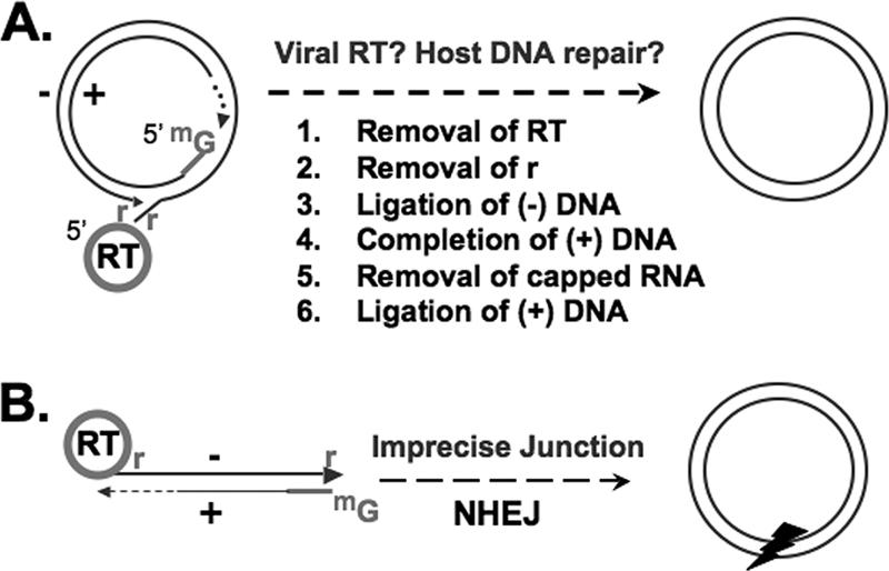
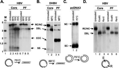
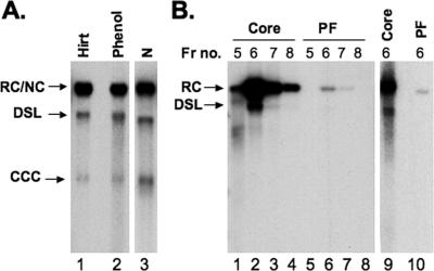
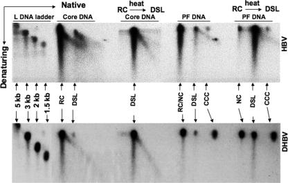
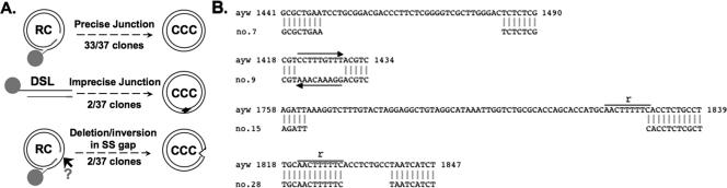
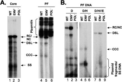
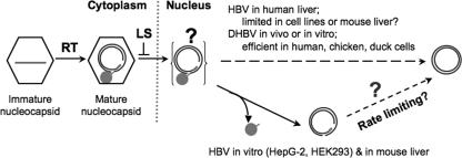
Hepatitis B virus (HBV) contains a small, partially double-stranded, relaxed circular (RC) DNA genome. RC DNA needs to be converted to covalently closed circular (CCC) DNA, which serves as the template for all viral RNA transcription. As a first step toward understanding how CCC DNA is formed, we analyzed the viral and host factors that may be involved in CCC DNA formation, using transient and stable DNA transfections of HBV and the related avian hepadnavirus, duck hepatitis B virus (DHBV). Our results show that HBV CCC DNA formed in hepatoma cells was derived predominantly from RC DNA with a precise junction sequence. In contrast to that of DHBV, HBV CCC DNA formation in cultured cells was accompanied by the accumulation of a RC DNA species from which the covalently attached viral reverse transcriptase (RT) protein was removed (protein-free or PF-RC DNA). Furthermore, whereas envelope deficiency led to increased CCC DNA formation in DHBV, it resulted mainly in increased PF-RC, but not CCC, DNA in HBV, suggesting that the envelope protein(s) may negatively regulate a step in CCC DNA formation that precedes deproteination in both HBV and DHBV. Interestingly, PF-RC DNA, in contrast to RT-linked RC DNA, contained, almost exclusively, mature plus-strand DNA, suggesting that the RT protein was removed preferentially from mature RC DNA.


Figures

FIG. 1.
FIG. 1.
Conversion of RC or DSL DNA to CCC DNA. (A) Structure of genomic RC DNA, which accounts for ca. 90% of reverse transcription products. Shown are the RT protein covalently attached to the 5′ end of the minus-strand DNA [(−) DNA], the r of the minus-strand DNA, and the capped RNA oligomer (denoted by methyl guanosine [mG]) attached to the 5′ end of the incomplete (gapped) plus-strand DNA [(+) DNA], with heterogeneous 3′ ends. Conversion of RC DNA to CCC DNA is a multistep process, as shown. (B) DSL DNA, with ends modified similar to those of RC DNA, except that the 5′ ends of the two strands are not annealed. DSL DNA accounts for ca. 10% of genomic DNA and is thought to be converted to CCC DNA via NHEJ, an imprecise process that results in deletions/insertions at the junction, denoted by an arrowhead.
FIG. 2.
FIG. 2.
Formation of HBV and DHBV CCC DNA in stably transfected hepatoma cell lines. HBV (A) and DHBV (B) DNAs were extracted from the cytoplasmic nucleocapsid (core) with protease digestion or the Hirt supernatant without protease digestion (PF), respectively, from HepAD38 (HBV) and Dstet5 (DHBV) cells cultured in the absence of Tet and analyzed by Southern blotting. The nature of CCC DNA in the HBV PF sample (panel A, lanes 5 and 6) was further confirmed by heating to 95°C, which denatured RC/NC and DSL DNA to SS without denaturing CCC DNA, or at 95°C, followed by EcoRI digestion, which linearized CCC DNA. Note the predominance of CCC DNA and RC/NC DNA in the DHBV and HBV PF DNAs, respectively. El, EcoRI. (C) The kinetics of accumulation of CCC DNA and PF-RC DNA in HepAD38 (HBV) and Dstet5 (DHBV) cells. The PF DNA was extracted after induction for 7, 10, 14, and 17 days for HBV (lanes 1 to 4) and 4, 5, 6, and 7 days for DHBV (lanes 5 to 8) and analyzed by Southern blotting.
FIG. 3.
FIG. 3.
Analyses of PF-RC DNA. HBV (A) and DHBV (B) core and PF DNA, extracted as for Fig. 2, or the plasmid pcDNA3 (C) were heated at the indicated temperatures and analyzed by Southern blotting. The diagrams at the bottom depict that only RC, not NC, is linearized at the indicated temperatures. SC, supercoiled plasmid. (D) HBV core or PF DNA was digested with the indicated restriction enzymes and analyzed by Southern blotting. The diagram at the bottom depicts the restriction sites on the RC DNA. E, EcoRI; R, RsrII.
FIG. 4.
FIG. 4.
Detection of HBV PF DNA in the nucleus and nucleocapsid. (A) HBV PF DNA was isolated from the whole-cell lysate, using either Hirt extraction (lane 1) or direct phenol extraction (lane 2), or from the nuclear fraction (N, lane 3) by Hirt extraction. (B) HBV DNA was isolated from the peak fractions of nucleocapsids purified by CsCl gradient centrifugation with protease digestion for core DNA (lanes 1 to 4) or without protease digestion for PF DNA (lanes 5 to 8). HBV core DNA (lane 9) and PF DNA (lane 10) were also extracted from the purified HBV nucleocapsids (fraction 6) after being mixed with the total HepG2 cell lysate. Fr no., fraction number.
FIG. 5.
FIG. 5.
Two-dimensional agarose gel electrophoresis analyses of core and PF DNAs. The HBV (top panel) and DHBV (bottom panel) core and PF DNAs were extracted as for Fig. 2. The DNA was separated first on a native agarose gel (first dimension), followed by a denaturing alkaline agarose gel (second dimension). After being transferred to a nylon membrane, viral plus-strand DNA was detected by using an antisense riboprobe. Linear DNA markers were run in parallel as size standards (left side). Where indicated, the DNA samples were heated (68°C for DHBV and 75°C for HBV) to convert RC to the DSL form. For clarity, only DNA species migrating above the full-length minus-strand SS DNA (Fig. 2) were analyzed on the second-dimension gel.
FIG. 6.
FIG. 6.
Sequencing results for CCC DNA at the cohesive-overlap region. The HBV CCC DNA was gel-purified from the PF DNA sample extracted from HepAD38 cells. The sequence spanning the cohesive overlap (Fig. 1) was amplified by PCR, cloned into the plasmid pcDNA3, and sequenced. (A) Schematic diagram summarizing the sequence results. The deletions detected in CCC DNA molecules derived from circularization of DSL DNA are denoted with an arrowhead. The other deletion and inversion events in the CCC DNA were detected in sequences corresponding to the presumed SS gap (arrow) of RC (and DSL, omitted for clarity) DNA. (B) Sequences of four individual clones that suffered deletions or inversion. The sequence on the top line is from the input WT HBV (strain ayw). The deletions and inversion in the sequences shown on the bottom line are indicated by blank spaces (deletion) or a left-pointing arrow (inversion). The nucleotide positions are indicated, as is the 9-nt r sequence. See the text for details.
FIG. 7.
FIG. 7.
HBV core DNA and PF DNA in transiently transfected HepG-2 cells. (A) pCMV-HBV (WT), pCMV-HBV/ENV, and pCMV-HBV/POL were transfected into HepG-2 cells. Viral core (lanes 1 to 3) and PF (lanes 4 to 6) DNA samples were harvested 5 days after transfection and analyzed by Southern blotting. The cells were transfected in duplicate, with one of the two dishes used for isolation of core DNA and the other for PF DNA. (B) PF DNA was further analyzed by DpnI digestion (lanes 1 to 3, D) to degrade input plasmids; DpnI digestion, followed by heating to 95°C (lanes 4 to 6, D/H), to denature the plasmid fragments, RC/NC and DSL DNA, but not CCC DNA, to SS DNA; or DpnI digestion, followed by heating and then EcoRI digestion (lanes 7 to 9, D/H/E), to convert CCC DNA to a linear form. The different viral DNA species, as well as the input plasmids, are indicated. This experiment was repeated three times with similar results.
FIG. 8.
FIG. 8.
Potential viral and host factors influencing RC DNA to CCC DNA conversion. Shown schematically are the immature (containing pgRNA or SS DNA) and mature (containing mature RC DNA) nucleocapsids in the cytoplasm, the hypothetical nuclear RC DNA intermediate still linked to the RT after uncoating (not yet detected), PF-RC DNA (detected in this report), and CCC DNA. Only mature RC DNA, not immature DNA, is shown to be deproteinated, possibly as a result of preferential uncoating of mature nucleocapsids and/or nuclear import of mature RC DNA, which is proposed to be regulated negatively by the viral LS protein. The predominance of HBV and DHBV CCC or RC DNA under different in vivo and in vitro conditions is indicated. RT, reverse transcription. See the text for details.

References

    1. Abdelhamed, A. M., C. M. Kelley, T. G. Miller, P. A. Furman, E. E. Cable, and H. C. Isom. 2003. Comparison of anti-hepatitis B virus activities of lamivudine and clevudine by a quantitative assay. Antimicrob. Agents Chemother. 47:324-336. - PMC - PubMed
    1. Acs, G., M. A. Sells, R. H. Purcell, P. Price, R. Engle, M. Shapiro, and H. Popper. 1987. Hepatitis B virus produced by transfected Hep G2 cells causes hepatitis in chimpanzees. Proc. Natl. Acad. Sci. USA 84:4641-4644. - PMC - PubMed
    1. Anderson, A. L., K. E. Banks, M. Pontoglio, M. Yaniv, and A. McLachlan. 2005. Alpha/beta interferon differentially modulates the clearance of cytoplasmic encapsidated replication intermediates and nuclear covalently closed circular hepatitis B virus (HBV) DNA from the livers of hepatocyte nuclear factor 1α-null HBV transgenic mice. J. Virol. 79:11045-11052. - PMC - PubMed
    1. Bartenschlager, R., and H. Schaller. 1992. Hepadnaviral assembly is initiated by polymerase binding to the encapsidation signal in the viral RNA genome. EMBO J. 11:3413-3420. - PMC - PubMed
    1. Bruss, V. 1997. A short linear sequence in the pre-S domain of the large hepatitis B virus envelope protein required for virion formation. J. Virol. 71:9350-9357. - PMC - PubMed

Publication types

Cite

AltStyle によって変換されたページ (->オリジナル) /