This site needs JavaScript to work properly. Please enable it to take advantage of the complete set of features!
Skip to main page content
U.S. flag

An official website of the United States government

Dot gov

The .gov means it’s official.
Federal government websites often end in .gov or .mil. Before sharing sensitive information, make sure you’re on a federal government site.

Https

The site is secure.
The https:// ensures that you are connecting to the official website and that any information you provide is encrypted and transmitted securely.

NIH NLM Logo
Log in
Access keys NCBI Homepage MyNCBI Homepage Main Content Main Navigation
. 2006 Feb;26(3):735-42.
doi: 10.1128/MCB.26.3.735-742.2006.

Tollip regulates proinflammatory responses to interleukin-1 and lipopolysaccharide

Affiliations

Tollip regulates proinflammatory responses to interleukin-1 and lipopolysaccharide

Arnaud Didierlaurent et al. Mol Cell Biol. 2006 Feb.

Abstract

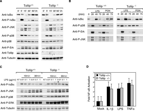
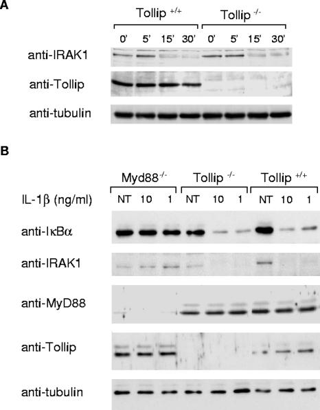
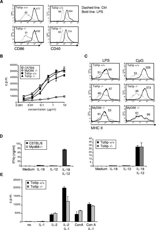
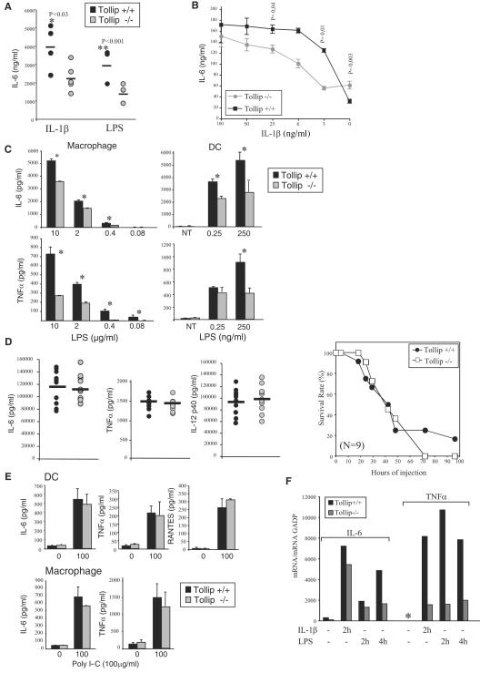
Activation of interleukin-1 (IL-1) receptor (IL-1R), Toll-like receptor 2 (TLR2), and TLR4 triggers NF-kappaB and mitogen-activated protein kinase (MAPK)-dependent signaling, thereby initiating immune responses. Tollip has been implicated as a negative regulator of NF-kappaB signaling triggered by these receptors in in vitro studies. Here, deficient mice were used to determine the physiological contribution of Tollip to immunity. NF-kappaB, as well as MAPK, signaling appeared normal in Tollip-deficient cells stimulated with IL-1beta or the TLR4 ligand lipopolysaccharide (LPS). Similarly, IL-1beta- and TLR-driven activation of dendritic cells and lymphocytes was indistinguishable from wild-type cells. In contrast, the production of the proinflammatory cytokines, IL-6 and tumor necrosis factor alpha was significantly reduced after IL-1beta and LPS treatment at low doses but not at lethal doses of LPS. Tollip therefore controls the magnitude of inflammatory cytokine production in response to IL-1beta and LPS.


Figures

FIG. 1.
FIG. 1.
Generation of Tollip−/− mice. (A) Organization of the mouse Tollip gene, targeting vector, and mutated allele. Filled boxes denote exons (1-6). The arrow and asterisks denote the translational initiator ATG and terminator TAA sequences, respectively. (B) Analysis of genomic DNA by PCR using primers spanning the regions indicated in panel A. (C) Western blot analysis of cellular extracts from Tollip+/+ and Tollip−/− MEFs or 293T cells transfected with VSV TollipΔN (amino acids 47 to 274) or full-length VSV Tollip using antibodies to Tollip (upper panel) or VSV (middle panel). To confirm equal loading the same blot was reprobed with antitubulin antibodies (lower panel).
FIG. 2.
FIG. 2.
Analysis of IL-1β-, LPS-, and PGN-triggered signaling cascades. (A) MEFs (A) or bone marrow-derived macrophages (B) from wild-type (+/+) and Tollip-deficient (−/−) mice were stimulated with LPS (1 μg/ml), PGN (10 μg/ml), or IL-1β (50 ng/ml) for the indicated times. Cell lysates were prepared and immunoblotted with various antibodies as indicated. Shown are representative experiments, repeated at least three times with similar results. (C) Peritoneal macrophages from wild-type (+/+) and Tollip-deficient (−/−) mice were stimulated with LPS at the indicated concentrations. Cell lysates were prepared and immunoblotted with the indicated antibodies. (D) MEFs were transfected with 500 ng of a pLuc (a NF-κB-dependent reporter plasmid) and 20 ng of phRLTK (Renilla luciferase). The cells were stimulated for 6 h with IL-1β (20 ng/ml), LPS (1 μg/ml), or TNF-α (100 ng/ml) and lysed, and the luciferase activities determined by using the dual-luciferase reporter assay system. Values shown are from data obtained in two transfection experiments. NT, nontreated.
FIG. 3.
FIG. 3.
IRAK degradation occurs normally in Tollip−/− cells. (A) Tollip-deficient (−/−) or wild-type (+/+) MEFs were stimulated with IL-1β (20 ng/ml) for the indicated times. Cell lysates were prepared and immunoblotted with anti-IRAK-1 or anti-IκBα antibodies. (B) Tollip-deficient (−/−), wild-type (+/+), or MyD88-deficient (−/−) MEFs were stimulated with the indicated concentrations of IL-1β for 15 min. Cell lysates were prepared and immunoblotted with various antibodies as indicated. As a loading control in these experiments, the same membrane was reprobed with antibodies against tubulin. NT, not treated.
FIG. 4.
FIG. 4.
TLR- and IL-1β-dependent activation of immune cells is not impaired in the absence of Tollip. (A) Mice were injected intravenously with 1 μg of LPS, and 6 h later splenic DCs were analyzed by flow cytometry for the indicated activation markers. Histograms represent data for F4/80−/low CD11c+ gated cells. (B) Splenocytes were incubated with LPS at the indicated concentrations, and proliferation was monitored 48 h later by pulsing the cells overnight with [3H]thymidine. (C) Splenic B cells were isolated by using B220+ MACS beads and incubated with 5 μg of LPS/ml or 10 μg of CpG/ml for 48 h. Surface expression of MHC II was then monitored by flow cytometry. (D) Splenocytes were plated in triplicate in medium containing 10 ng of IL-12/ml, 20 ng of IL-18/ml, or both. The production of IFN-γ in the supernatant was measured by ELISA 72 h later. (E) Thymocytes were incubated in medium containing 20 U of IL-2/ml, 10 ng of IL-1β/ml, and 2.5 μg of ConA/ml as indicated for 3 days. The cell proliferation was measured by pulsing the cells overnight with [3H]thymidine. The data are averages of quadruplicates and are representative of three independent experiments.
FIG. 5.
FIG. 5.
Tollip-deficient mice produce less IL-6 and TNF-α mRNA and protein upon LPS and IL-1β stimulation. (A) Tollip−/− or wild-type mice were injected intravenously with 1 μg of IL-1β (five mice/group) or 1 μg of LPS (three mice/group), and the production of IL-6 in the serum was measured at 2 h by ELISA. (B) BMDM were treated with 100 ng of IFN-γ/ml and stimulated with the indicated concentrations of IL-1β, and the production of IL-6 in the supernatant was measured by ELISA 24 h later. (C) BMDM or BMDC were stimulated with the indicated concentrations of LPS, and the production of IL-6 and TNF-α in the supernatant was measured by ELISA 24 h later. The experiments in panels B and C were repeated at least twice in triplicates with similar results. Significant differences are indicated by an asterisks (✽, P < 0.04). (D) Tollip-deficient (−/−) or wild-type mice (+/+) were injected intraperitoneally with a lethal dose of LPS (E. coli O111:B4) at a dose of 40 mg/kg (body weight) to induce septic shock. The serum concentrations of IL-6, TNF-α, and IL-12p40 for all mice/group (n = 9) were measured by ELISA 3 h after injection, and the data shown are averages for each group. Animals were then monitored every 12 h for moribund state and then sacrificed. (E) Peritoneal macrophages or DC were stimulated with 10 or 100 μg of poly(I-C)/ml, and the production of IL-6, TNF-α, and RANTES in the supernatant was measured by ELISA 24 h later. DC from wild-type (+/+) and Tollip-deficient (−/−) mice were stimulated with LPS (1 μg/ml) or poly(I-C) (100 μg/ml) for 24 h. Cell lysates were prepared and immunoblotted as indicated. (F) Wild-type and Tollip−/− MEFs were stimulated with IL-1β (5 ng/ml) or LPS (100 ng/ml) for the indicated times, and the IL-6 or TNF-α mRNA levels were determined by quantitative real-time RT-PCR analysis. ✽, not detected.

References

    1. Akira, S., and K. Takeda. 2004. Toll-like receptor signalling. Nat. Rev. Immunol. 4:499-511. - PubMed
    1. Bulut, Y., E. Faure, L. Thomas, O. Equils, and M. Arditi. 2001. Cooperation of Toll-like receptor 2 and 6 for cellular activation by soluble tuberculosis factor and Borrelia burgdorferi outer surface protein A lipoprotein: role of Toll-interacting protein and IL-1 receptor signaling molecules in Toll-like receptor 2 signaling. J. Immunol. 167:987-994. - PubMed
    1. Burns, K., J. Clatworthy, L. Martin, F. Martinon, C. Plumpton, B. Maschera, A. Lewis, K. Ray, J. Tschopp, and F. Volpe. 2000. Tollip, a new component of the IL-1RI pathway, links IRAK to the IL-1 receptor. Nat. Cell Biol. 2:346-351. - PubMed
    1. Burns, K., S. Janssens, B. Brissoni, N. Olivos, R. Beyaert, and J. Tschopp. 2003. Inhibition of interleukin 1 receptor/Toll-like receptor signaling through the alternatively spliced, short form of MyD88 is due to its failure to recruit IRAK-4. J. Exp. Med. 197:263-268. - PMC - PubMed
    1. Didierlaurent, A., I. Ferrero, L. A. Otten, B. Dubois, M. Reinhardt, H. Carlsen, R. Blomhoff, S. Akira, J. P. Kraehenbuhl, and J. C. Sirard. 2004. Flagellin promotes myeloid differentiation factor 88-dependent development of Th2-type response. J. Immunol. 172:6922-6930. - PubMed

Publication types

MeSH terms

LinkOut - more resources

Cite

AltStyle によって変換されたページ (->オリジナル) /