This site needs JavaScript to work properly. Please enable it to take advantage of the complete set of features!
Skip to main page content
U.S. flag

An official website of the United States government

Dot gov

The .gov means it’s official.
Federal government websites often end in .gov or .mil. Before sharing sensitive information, make sure you’re on a federal government site.

Https

The site is secure.
The https:// ensures that you are connecting to the official website and that any information you provide is encrypted and transmitted securely.

NIH NLM Logo
Log in
Access keys NCBI Homepage MyNCBI Homepage Main Content Main Navigation
. 2005 Dec 2;33(21):6769-81.
doi: 10.1093/nar/gki1002. Print 2005.

The interaction site of Flap Endonuclease-1 with WRN helicase suggests a coordination of WRN and PCNA

Affiliations

The interaction site of Flap Endonuclease-1 with WRN helicase suggests a coordination of WRN and PCNA

Sudha Sharma et al. Nucleic Acids Res. .

Abstract

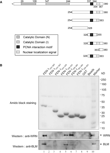
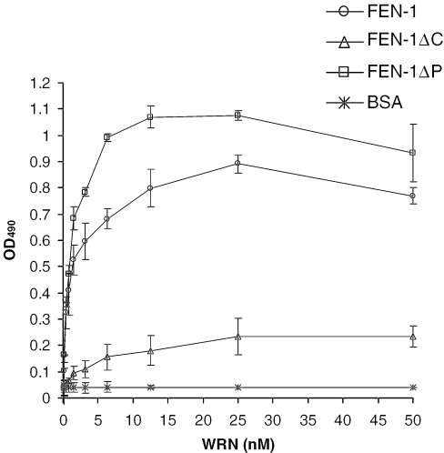
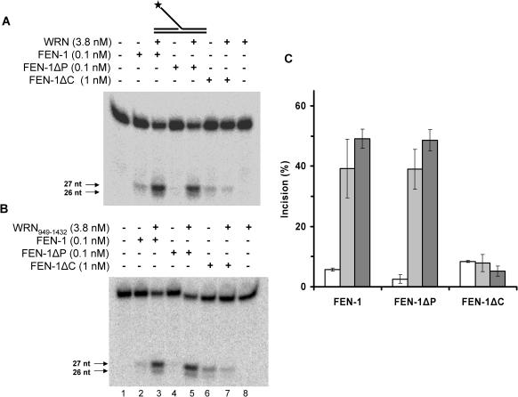
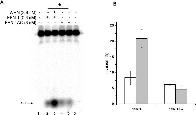
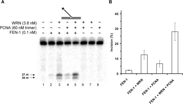
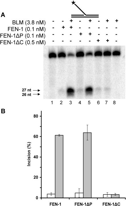
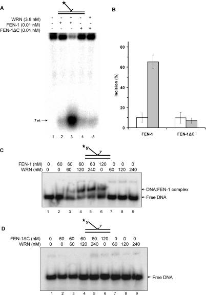
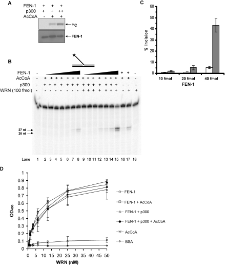
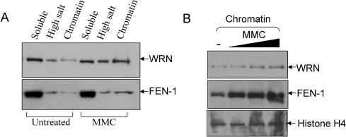
Werner and Bloom syndromes are genetic RecQ helicase disorders characterized by genomic instability. Biochemical and genetic data indicate that an important protein interaction of WRN and Bloom syndrome (BLM) helicases is with the structure-specific nuclease Flap Endonuclease 1 (FEN-1), an enzyme that is implicated in the processing of DNA intermediates that arise during cellular DNA replication, repair and recombination. To acquire a better understanding of the interaction of WRN and BLM with FEN-1, we have mapped the FEN-1 binding site on the two RecQ helicases. Both WRN and BLM bind to the extreme C-terminal 18 amino acid tail of FEN-1 that is adjacent to the PCNA binding site of FEN-1. The importance of the WRN/BLM physical interaction with the FEN-1 C-terminal tail was confirmed by functional interaction studies with catalytically active purified recombinant FEN-1 deletion mutant proteins that lack either the WRN/BLM binding site or the PCNA interaction site. The distinct binding sites of WRN and PCNA and their combined effect on FEN-1 nuclease activity suggest that they may coordinately act with FEN-1. WRN was shown to facilitate FEN-1 binding to its preferred double-flap substrate through its protein interaction with the FEN-1 C-terminal binding site. WRN retained its ability to physically bind and stimulate acetylated FEN-1 cleavage activity to the same extent as unacetylated FEN-1. These studies provide new insights to the interaction of WRN and BLM helicases with FEN-1, and how these interactions might be regulated with the PCNA-FEN-1 interaction during DNA replication and repair.


Figures

Figure 1
Figure 1
WRN or BLM binding activity of FEN-1 is contained within amino acids 363–380. (A) Schematic representation of GST-FEN-1 recombinant fragments used for WRN pull-down experiments. (B) Amido black-stained membrane of protein complexes bound to glutathione–agarose beads in pull-down binding assay. Beads were mixed with lysate from bacteria expressing GST fusion proteins containing human FEN-1 amino acids 254–380 (lane 1), 254–363 (lane 2), 254–328 (lane 3), 328–355 (lane 4), 328–380 (lane 5), 363–380 (lane 6), 328–363 (lane 7), GST alone (lane 8) or the agarose beads (lane 9). Purified recombinant WRN or BLM proteins (200 ng) were added to the indicated affinity beads. After washing, protein complexes were eluted and analyzed by SDS–PAGE. Bound WRN (upper panel) or BLM (lower panel) was detected by western blot analysis as described in Materials and Methods. Purified recombinant WRN or BLM protein (50 ng) was used as a marker for western blot (lane 10). In other experiments, greater amounts of the fusion protein GST–FEN-1254–363 bound to glutathione beads that were more similar to the other GST fusion proteins did not pull down WRN or BLM (data not shown).
Figure 2
Figure 2
Detection of WRN–FEN-1 complexes by ELISA. Wild-type FEN-1, FEN-1ΔC or FEN-1ΔP was coated onto ELISA plates. Following blocking with 3% BSA, the wells were incubated with increasing concentrations of purified recombinant WRN (0–50 nM) for 1 h at 30°C, and bound WRN was detected by ELISA using a rabbit polyclonal antibody against WRN followed by incubation with secondary HRP-labeled antibodies and OPD substrate. Data points are the mean of three independent experiments performed in duplicate with SDs indicated by error bars.
Figure 3
Figure 3
WRN-interacting region of FEN-1 is required for WRN stimulation of FEN-1 endonucleolytic cleavage of 5′ flap substrate. Reaction mixtures (20 μl) containing 10 fmol of the 26 nt 5′ flap substrate, the specified concentrations of wild-type FEN-1, FEN-1ΔC or FEN-1ΔP, and either WRN (A) or WRN949–1432 (B) as indicated were incubated at 37°C for 15 min under standard conditions as described in Materials and Methods. Products were resolved on 20% polyacrylamide denaturing gels. Phosphorimages of typical gels are shown. For each gel: lane 1, no enzyme; lanes 2, 4 and 6 are wild-type FEN-1, FEN-1ΔP and FEN-1ΔC, respectively; lanes 3, 5 and 7 are wild-type FEN-1, FEN-1ΔP and FEN-1ΔC, respectively, in the presence of WRN (A) or WRN949–1432 (B); lane 8, WRN (A) or WRN949–1432 (B) alone. (C) Per cent incision (mean value of at least three independent experiments with SDs indicated by error bars). Quantitative data are shown for incision reactions with FEN-1, FEN-1ΔP or FEN-1ΔC alone (open bars), in the presence of WRN (light gray bars) or in the presence of WRN949–1432 (gray bars).
Figure 4
Figure 4
WRN-interacting region of FEN-1 is required for WRN stimulation of FEN-1 exonucleolytic cleavage of nicked duplex substrate. Reaction mixtures (20 μl) containing 10 fmol of the nicked duplex DNA substrate, the specified concentrations of wild-type FEN-1 or FEN-1ΔC, and WRN as indicated were incubated at 37°C for 15 min under standard conditions as described in Materials and Methods. Products were resolved on 20% polyacrylamide denaturing gels. (A) Phosphorimage of a typical gel is shown. Lane 1, no enzyme; lanes 2 and 4 are wild-type FEN-1 and FEN-1ΔC, respectively; lanes 3 and 5 are wild-type FEN-1 and FEN-1ΔC, respectively, in the presence of WRN. (B) Per cent incision (mean value of at least three independent experiments with SD indicated by error bars). Quantitative data are shown for incision reactions with FEN-1 or FEN-1ΔC alone (open bars) or in the presence of WRN (light gray bars).
Figure 5
Figure 5
Combined effect of WRN and PCNA on stimulation of FEN-1 cleavage. Reaction mixtures (20 μl) containing 10 fmol of the 26 nt 5′ flap substrate and the indicated concentrations of FEN-1, WRN and/or PCNA were incubated at 37°C for 15 min under standard conditions as described in Materials and Methods. Products were resolved on 20% polyacrylamide denaturing gels. (A) Phosphorimage of a typical gel is shown. Lane 1, no enzyme; lane 2, FEN-1; lane 3, FEN-1 + WRN; lane 4, FEN-1 + PCNA; lane 5, FEN-1 + WRN + PCNA; lane 6, WRN + PCNA; lane 7, WRN; lane 8, PCNA. (B) Per cent incision (mean value of at least three independent experiments with SD indicated by error bars).
Figure 6
Figure 6
BLM-interacting region of FEN-1 is required for BLM stimulation of FEN-1 endonucleolytic cleavage of 5′ flap substrate. Reaction mixtures (20 μl) containing 10 fmol of the 26 nt 5′ flap substrate, the specified concentrations of wild-type FEN-1, FEN-1ΔC or FEN-1ΔP, and BLM were incubated at 37°C for 15 min under standard conditions as described in Materials and Methods. Products were resolved on 20% polyacrylamide denaturing gels. (A) Phosphorimage of a typical gel is shown. Lane 1, no enzyme; lanes 2, 4 and 6 are wild-type FEN-1, FEN-1ΔP and FEN-1ΔC, respectively; lanes 3, 5 and 7 are wild-type FEN-1, FEN-1ΔP and FEN-1ΔC, respectively, in the presence of BLM; lane 8, BLM alone. (B) Per cent incision (mean value of at least three independent experiments with SD indicated by error bars). Quantitative data are shown for incision reactions with FEN-1 or FEN-1ΔC alone (open bars) or in the presence of BLM (light gray bars).
Figure 7
Figure 7
WRN facilitates FEN-1 binding to the double-flap DNA substrate through its interaction with the C-terminal tail of FEN-1. (A and B) Reaction mixtures (20 μl) containing 10 fmol of the double flap DNA substrate, the specified concentrations of wild-type FEN-1 or FEN-1ΔC, and WRN as indicated were incubated at 37°C for 15 min under standard conditions as described in Materials and Methods. Products were resolved on 20% polyacrylamide denaturing gels. (A) Phosphorimage of a typical gel is shown. Lane 1, no enzyme; lanes 2 and 4 are wild-type FEN-1 and FEN-1ΔC, respectively; lanes 3 and 5 are wild-type FEN-1 and FEN-1ΔC, respectively, in the presence of WRN. (B) Per cent incision (mean value of at least three independent experiments with SDs indicated by error bars). Quantitative data are shown for incision reactions with FEN-1 or FEN-1ΔC alone (open bars) or in the presence of WRN (light gray bars). (C and D) Binding mixtures (20 μl) containing 10 fmol of the double flap DNA substrate, the specified concentrations of wild-type FEN-1 (C) or FEN-1ΔC (D), and WRN as indicated were incubated at 37°C for 15 min as described in Materials and Methods. Products were resolved on native 5% polyacrylamide gels. Phosphorimages of typical gels are shown.
Figure 8
Figure 8
WRN retains the ability to physically bind and stimulate acetylated FEN-1. (A) Purified recombinant FEN-1 was acetylated in vitro by incubating with p300-HAT and [14C]acetyl coenzyme A (AcCoA) as described in Materials and Methods and resolved on SDS denaturing 10% polyacrylamide gel. The gel was exposed to X-ray film and developed by autoradiography (upper panel) or stained with Coomassie (lower panel). (B) Reaction mixtures (20 μl) containing 10 fmol of the 26 nt 5′ flap substrate, acetylated or unacetylated FEN-1 (0.625, 1.25, 2.5, 5, 10, 20 and 40 fmol) and WRN (100 fmol) were incubated at 37°C for 15 min under standard conditions as described in Materials and Methods. Products were resolved on 20% polyacrylamide denaturing gels. A phosphorimage of a typical gel is shown. Lanes 16 and 17 show the products of cleavage reactions containing 0.625 fmol FEN-1 (with AcCoA but lacking p300) in the absence and presence of WRN, respectively. (C) Per cent incision (mean value of at least three independent experiments with SD indicated by error bars from experiments as conducted in (B). Quantitative data are shown for incision reactions with acetylated FEN-1 alone (open bars) or in the presence of WRN (light gray bars). (D) FEN-1 that had been preincubated with or without p300-HAT and/or AcCoA as indicated was coated onto ELISA plates. Following blocking with 3% BSA, the wells were incubated with increasing concentrations of purified recombinant WRN (0–50 nM) for 1 h at 30°C, and bound WRN was detected by ELISA using a rabbit polyclonal antibody against WRN followed by incubation with secondary HRP-labeled antibodies and OPD substrate. Data points are the mean of three independent experiments performed in duplicate with SDs indicated by error bars.
Figure 9
Figure 9
Enriched association of WRN–FEN-1 in the chromatin fraction after DNA damage. (A) Greater fraction of WRN and FEN-1 is found in the chromatin fraction after exposure of cells to MMC. Subnuclear fractionation was performed as described in Materials and Methods. Western blot detection of WRN and FEN-1 in different subnuclear fractions of HeLa cells treated with or without MMC (0.5 μg/ml). (B) Enrichment of WRN and FEN-1 in the chromatin fraction is dependent on MMC dose. Western blot detection of WRN and FEN-1 in the chromatin fractions prepared from HeLa cells treated with or without 0.25, 0.5 or 1 μg/ml MMC. Bottom panel shows Histone H4 detected by western blot.

References

    1. Martin G.M. Genetic syndromes in man with potential relevance to the pathobiology of aging. Birth Defects Orig. Artic. Ser. 1978;14:5–15. - PubMed
    1. Hanaoka F., Yamada M., Takeuchi F., Goto M., Miyamoto T., Hori T. Autoradiographic studies of DNA replication in Werner's syndrome cells. Adv. Exp. Med. Biol. 1985;190:439–457. - PubMed
    1. Martin G.M., Sprague C.A., Epstein C.J. Replicative life-span of cultivated human cells. Effects of donor's age, tissue, and genotype. Lab. Invest. 1970;23:86–92. - PubMed
    1. Poot M., Hoehn H., Runger T.M., Martin G.M. Impaired S-phase transit of Werner syndrome cells expressed in lymphoblastoid cells. Exp. Cell. Res. 1992;202:267–273. - PubMed
    1. Prince P.R., Emond M.J., Monnat R.J., Jr Loss of Werner syndrome protein function promotes aberrant mitotic recombination. Genes Dev. 2001;15:933–938. - PMC - PubMed

Publication types

Cite

AltStyle によって変換されたページ (->オリジナル) /