This site needs JavaScript to work properly. Please enable it to take advantage of the complete set of features!
Skip to main page content
U.S. flag

An official website of the United States government

Dot gov

The .gov means it’s official.
Federal government websites often end in .gov or .mil. Before sharing sensitive information, make sure you’re on a federal government site.

Https

The site is secure.
The https:// ensures that you are connecting to the official website and that any information you provide is encrypted and transmitted securely.

NIH NLM Logo
Log in
Access keys NCBI Homepage MyNCBI Homepage Main Content Main Navigation
. 2005 Jul;79(13):8004-13.
doi: 10.1128/JVI.79.13.8004-8013.2005.

Inhibition of alpha/beta interferon signaling by the NS4B protein of flaviviruses

Affiliations

Inhibition of alpha/beta interferon signaling by the NS4B protein of flaviviruses

Jorge L Muñoz-Jordán et al. J Virol. 2005 Jul.

Abstract

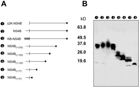
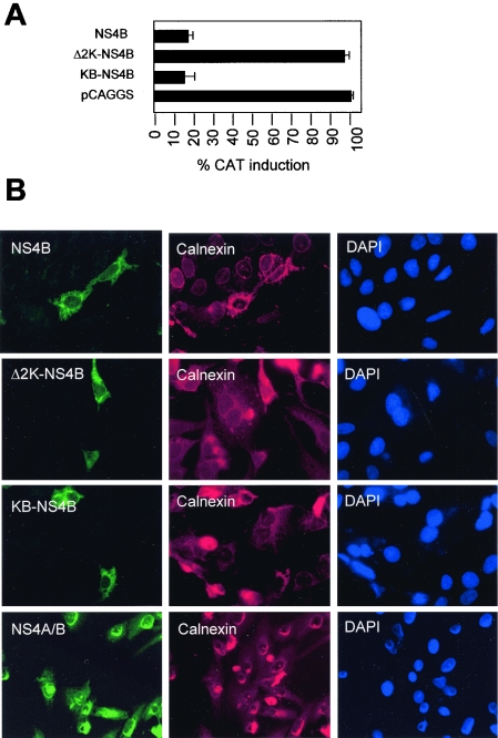
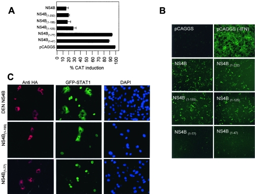
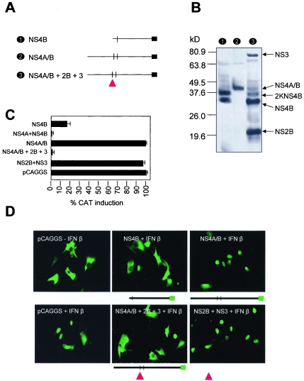
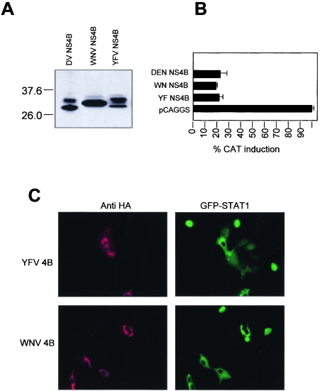
Flaviviruses are insect-borne, positive-strand RNA viruses that have been disseminated worldwide. Their genome is translated into a polyprotein, which is subsequently cleaved by a combination of viral and host proteases to produce three structural proteins and seven nonstructural proteins. The nonstructural protein NS4B of dengue 2 virus partially blocks activation of STAT1 and interferon-stimulated response element (ISRE) promoters in cells stimulated with interferon (IFN). We have found that this function of NS4B is conserved in West Nile and yellow fever viruses. Deletion analysis shows that that the first 125 amino acids of dengue virus NS4B are sufficient for inhibition of alpha/beta IFN (IFN-alpha/beta) signaling. The cleavable signal peptide at the N terminus of NS4B, a peptide with a molecular weight of 2,000, is required for IFN antagonism but can be replaced by an unrelated signal peptide. Coexpression of dengue virus NS4A and NS4B together results in enhanced inhibition of ISRE promoter activation in response to IFN-alpha/beta. In contrast, expression of the precursor NS4A/B fusion protein does not cause an inhibition of IFN signaling unless this product is cleaved by the viral peptidase NS2B/NS3, indicating that proper viral polyprotein processing is required for anti-interferon function.


Figures

FIG. 1.
FIG. 1.
Deletion analysis of DEN NS4B. (A) Schematic representation of various deletions of DEN-2 NS4B created by PCR. The N-terminal 2K segment was deleted (Δ2K-NS4B), left intact (NS4B), or replaced by the murine MHC-I signal peptide KB (KB-NS4B). In addition, deletions of the C-terminal region were also created by PCR [NS4B(1-232), NS4B(1-155), NS4B(1-125), NS4B(1-77), and NS4B(1-47)] as indicated. (B) Immunoblot analysis of HA-tagged NS4B derivatives. All fragments represented in panel A were cloned in pCAGGS-HA to tag the C-terminal end of each protein. Expression levels of each protein were examined by running transfected cell extracts in a 4 to 20% gradient polyacrylamide. Proteins were transferred to a nitrocellulose membrane and detected with anti-HA primary antibody and horseradish peroxidase-labeled secondary antibody. Molecular mass (kDa) markers are at left.
FIG. 2.
FIG. 2.
Expression of 2K-mutant NS4Bs. (A) Induction of ISRE-9-27-CAT reporter gene after treatment with IFN. Recombinant pCAGGS-HA plasmids containing the DEN-2 NS4B, Δ2K-NS4B, or KB-NS4B gene fragments were transfected in Vero cells along with reporter plasmid ISRE-9-27-CAT and pCAGGS-FL. Following stimulation with 1,000 U of human IFN-β, CAT activity was quantitated in crude cell extracts. CAT activities were normalized to the corresponding FL activities to determine the percentage of CAT induction. CAT activities were determined as mean values from three independent experiments (P values of 0.02 to 0.06) (B) Immunostaining of transfected Vero cells. The indicated NS4B derivatives were expressed in Vero cells for 24 h. Cells were fixed, permeabilized, and immunostained using a polyclonal primary antibody for HA and a monoclonal anti-calnexin antibody. Fluorescein isothiocyanate-labeled secondary anti-rabbit antibody was used to detect NS4B proteins (green fluorescence) and a Texas red-labeled monoclonal antibody was used as a secondary antibody to detect calnexin (red fluorescence). DAPI staining reveals nuclear chromatin.
FIG. 3.
FIG. 3.
Analysis of C-terminal deletions of DEN NS4B. (A) Induction of ISRE-9-27-CAT reporter gene after treatment with IFN. Recombinant pCAGGS-HA plasmids containing each of the DEN-2 NS4B C-terminal deletions were transfected in Vero cells together with the ISRE-9-27-CAT plasmid, and CAT expression was stimulated with 1,000 U of IFN-β 24 h later. Results show the percentage of CAT activity for each individual construct. CAT activities were determined as mean values from two independent experiments (P values of 0.01 to 0.05). (B) Inhibition of NDV-GFP replication by IFN in the presence of DEN-2 NS4B derivatives. Vero cells were transfected with the plasmids expressing the indicated NS4B wild-type and mutant proteins and stimulated with 1,000 U of IFN-β prior to infection with NDV-GFP. Expression of GFP was visualized as green fluorescence by fluorescence microscopy. (C) STAT1 activation by IFN in the presence of NS4B derivatives. Vero cells simultaneously expressing NS4B (red) derivatives and GFP-STAT1 (green) were stimulated with 1,000 U of human IFN-β for 35 min, fixed, and permeabilized. HA-labeled protein was detected by fluorescence microscopy after incubation with polyclonal antibodies to HA and Texas red-labeled secondary antibody.
FIG. 4.
FIG. 4.
Cleavage of NS4A/B. (A) Schematic representation of NS4A/B constructs. NS4A/B was derived by PCR and cloned in pCAGGS-HA. A schematic representation shows NS4B, NS4A/B, and NS4A/B in the presence of NS2B and NS3 (indicated by a triangle). (B) An immunoblot shows expression of these recombinant proteins in Vero cells. In the presence of NS2B and NS3, a complex pattern is obtained with bands coinciding with those obtained when only NS4B is transfected as indicated. Molecular mass markers (kDa) are at left. (C) Induction of ISRE-9-27-CAT after treatment with IFN. The percentage of CAT induction by IFN-β in the presence of the indicated expression plasmids is shown. CAT activities were determined as mean values from two independent experiments (P values of 0.03 to 0.05). (D) Activation of STAT1 by IFN in the presence of cleaved and uncleaved DEN proteins. GFP-tagged STAT1 protein was visualized by fluorescence microscopy in cells previously transfected with the indicated constructs and briefly stimulated with IFN-β. NS4A/B+2B+3, NS4A/B+NS2B+NS3. The presence of NS2B+NS3 is indicated by a triangle.
FIG. 5.
FIG. 5.
Conserved NS4B function among flaviviruses. (A) An immunoblot shows levels of expression of DEN, WNV, and YFV NS4B in Vero cells transfected with the indicated plasmids. (B) Induction of ISRE-9-27-CAT after treatment with IFN-β. Vero cells were transfected with each of the indicated plasmids. At 24 h posttransfection, cells were treated with 1,000 U of human IFN-β. The CAT activities were normalized to the corresponding luciferase activities to determine CAT induction. CAT activities were determined as mean values from two independent experiments (P values of 0.02 to 0.04). (C) Vero cells simultaneously expressing YFV or WNV NS4B (red) and GFP-STAT1 (green) were treated with IFN-β. HA-labeled protein was detected by fluorescence microscopy after incubation with polyclonal antibodies to HA and Texas red-labeled secondary antibody.

References

    1. Amberg, S., A. Nestorowicz, D. McCourt, and C. Rice. 1994. NS2B-3 proteinase-mediated processing in the yellow fever virus structural region: in vitro and in vivo studies. J. Virol. 68:3794-3802. - PMC - PubMed
    1. Cahour, A., and B. Falgout. 1992. Cleavage of the dengue virus polyprotein at the NS3/NS4A and NS4A/NS5 junctions is mediated by viral protease NS2B-NS3, whereas NS4A/NS4B may be processed by a cellular protease. J. Virol. 66:1535-1542. - PMC - PubMed
    1. Chen, Y., T. Maguire, and R. Marks. 1996. Demonstration of binding of dengue virus envelope protein to target cells. J. Virol. 70:8765-8772. - PMC - PubMed
    1. Chu, P. W., and E. G. Westaway. 1992. Molecular and ultrastructural analysis of heavy membrane fractions associated with the replication of Kunjin virus RNA. Arch. Virol. 125:177-191. - PubMed
    1. Deblandre, G. A., O. P. Marinx, S. S. Evans, S. Majjaj, O. Leo, D. Caput, G. A. Huez, and M. G. Wathelet. 1995. Expression cloning of an interferon-inducible 17-kDa membrane protein implicated in the control of cell growth. J. Biol. Chem. 270:23860-23866. - PubMed

Publication types

LinkOut - more resources

Cite

AltStyle によって変換されたページ (->オリジナル) /