This site needs JavaScript to work properly. Please enable it to take advantage of the complete set of features!
Skip to main page content
U.S. flag

An official website of the United States government

Dot gov

The .gov means it’s official.
Federal government websites often end in .gov or .mil. Before sharing sensitive information, make sure you’re on a federal government site.

Https

The site is secure.
The https:// ensures that you are connecting to the official website and that any information you provide is encrypted and transmitted securely.

NIH NLM Logo
Log in
Access keys NCBI Homepage MyNCBI Homepage Main Content Main Navigation
Review
. 2004 Apr;17(2):434-64.
doi: 10.1128/CMR.17.2.434-464.2004.

Intraspecific diversity of Yersinia pestis

Affiliations
Review

Intraspecific diversity of Yersinia pestis

Andrey P Anisimov et al. Clin Microbiol Rev. 2004 Apr.

Erratum in

  • Clin Microbiol Rev. 2004 Jul;17(3):695

Abstract

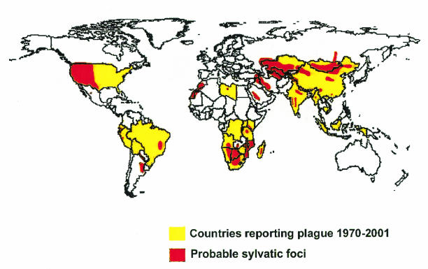
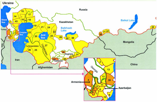
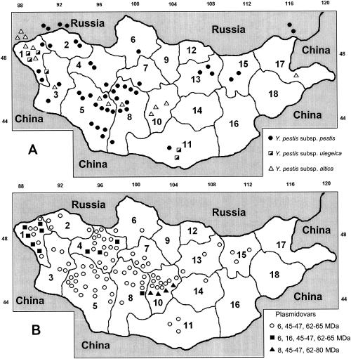
Increased interest in the pathogenic potential of Yersinia pestis has emerged because of the potential threats from bioterrorism. Pathogenic potential is based on genetic factors present in a population of microbes, yet most studies evaluating the role of specific genes in virulence have used a limited number of strains. For Y. pestis this issue is complicated by the fact that most strains available for study in the Americas are clonally derived and thus genetically restricted, emanating from a strain of Y. pestis introduced into the United States in 1902 via marine shipping and subsequent spread of this strain throughout North and South America. In countries from the former Soviet Union (FSU), Mongolia, and China there are large areas of enzootic foci of Y. pestis infection containing genetically diverse strains that have been intensely studied by scientists in these countries. However, the results of these investigations are not generally known outside of these countries. Here we describe the variety of methods used in the FSU to classify Y. pestis strains based on genetic and phenotypic variation and show that there is a high level of diversity in these strains not reflected by ones obtained from sylvatic areas and patients in the Americas.


Figures

FIG. 1.
FIG. 1.
Global distribution of plague. Reprinted with permission from K. L. Gage and J. A. Montenieri, Centers for Disease Control and Prevention, Fort Collins, Colo.
FIG. 2.
FIG. 2.
Distribution of natural plague foci in the FSU. Identifications of the plague foci are given in Table 1 and the text. The red line indicates the FSU frontier; green lines indicate the frontiers of the states of the FSU; brown lines indicate boundaries of the plague foci; black lines indicate frontiers of other countries. Plague foci are yellow (for foci containing the main Y. pestis subspecies) or light brown (for foci containing non-main Y. pestis subspecies). This figure is based on references , , , , and .
FIG. 3.
FIG. 3.
Distribution of Y. pestis subspecies (A) and plasmidovars (B) in Mongolia. Symbols for provinces (aymags): 1, Bayanölgie; 2, Uvs; 3, Hovd; 4, Dzavham; 5, Govï-Altay; 6, Hovsgol; 7, Arhangay; 8, Bayanhongor; 9, Bulgan; 10, Övörhangay; 11, Ömnögovï; 12, Selenge; 13, Töv; 14, Dundgovï; 15, Hentiy; 16, Dornogovï; 17, Dornod; 18, Sühbaatar. Panel A reprinted with permission from S. V. Balakhonov (15), Antiplague Research Institute of Siberia and Far East, Irkutsk, Russia. Panel B reprinted with permission from A. Erdenebat (62), Centre for Control and Research of Natural Infectious Diseases, Ulaanbaatar, Mongolia.
FIG. 4.
FIG. 4.
Distribution of Y. pestis plasmidovars in some Central Asian natural foci. Reprinted with permission from S. V. Balakhonov (17), Antiplague Research Institute of Siberia and Far East, Irkutsk, Russia.

References

    1. Abgaryan, G. P. 1966. Characterization of some Yersinia pestis strains, which were isolated on Armenian Highland from common voles. Ph.D. thesis. All-Union Research Anti-Plague Institute "Microbe," Saratov, USSR.
    1. Achtman, M., K. Zurth, G. Morelli, G. Torrea, A. Guiyoule, and E. Carniel. 1999. Yersinia pestis, the cause of plague, is a recently emerged clone of Yersinia pseudotuberculosis. Proc. Natl. Acad. Sci. USA 96:14043-14048. (Erratum, 97:8192, 2000.) - PMC - PubMed
    1. Adair, D. M., P. L. Worsham, K. K. Hill, A. M. Klevytska, P. J. Jackson, A. M. Friedlander, and P. Keim. 2000. Diversity in a variable-number tandem repeat from Yersinia pestis. J. Clin. Microbiol. 38:1516-1519. - PMC - PubMed
    1. Anderson, G. W. Jr., D. G. Heath, C. R. Bolt, S. L. Welkos, A. M. Friedlander. 1998. Short- and long-term efficacy of single-dose subunit vaccines against Yersinia pestis in mice. Am. J. Trop. Med. Hyg. 58:793-799. - PubMed
    1. Anderson, G. W., S. E. C. Leary, E. D. Williamson, R. W. Titball, S. L. Welkos, P. L. Worsham, and A. M. Friedlander. 1996. Recombinant V antigen protects mice against pneumonic and bubonic plague caused by F1-capsule-positive and F1-capsule-negative strains of Yersinia pestis. Infect. Immun. 64:4580-4585. - PMC - PubMed

Publication types

LinkOut - more resources

Cite

AltStyle によって変換されたページ (->オリジナル) /