This site needs JavaScript to work properly. Please enable it to take advantage of the complete set of features!
Skip to main page content
U.S. flag

An official website of the United States government

Dot gov

The .gov means it’s official.
Federal government websites often end in .gov or .mil. Before sharing sensitive information, make sure you’re on a federal government site.

Https

The site is secure.
The https:// ensures that you are connecting to the official website and that any information you provide is encrypted and transmitted securely.

NIH NLM Logo
Log in
Access keys NCBI Homepage MyNCBI Homepage Main Content Main Navigation
. 2004 Apr;72(4):1920-8.
doi: 10.1128/IAI.72.4.1920-1928.2004.

Toll-like receptor 4 contributes to efficient control of infection with the protozoan parasite Leishmania major

Affiliations

Toll-like receptor 4 contributes to efficient control of infection with the protozoan parasite Leishmania major

Pascale Kropf et al. Infect Immun. 2004 Apr.

Abstract

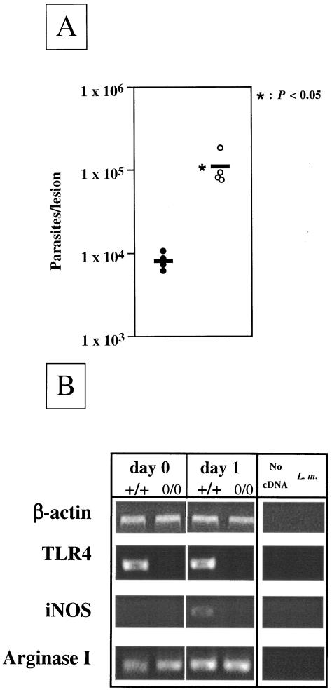
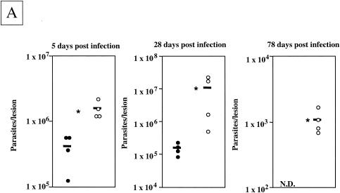
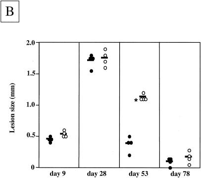
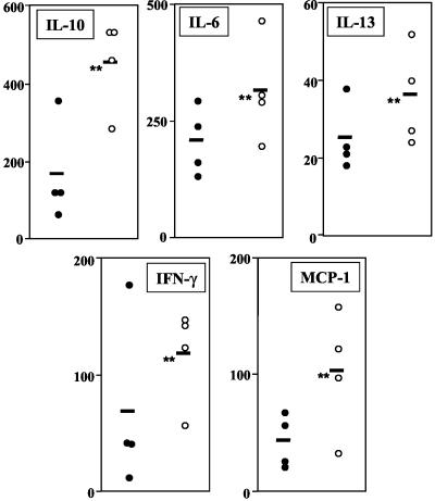
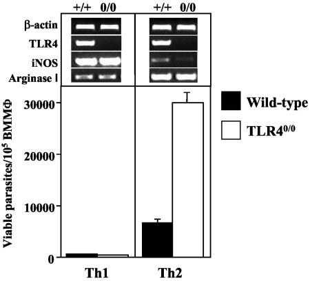
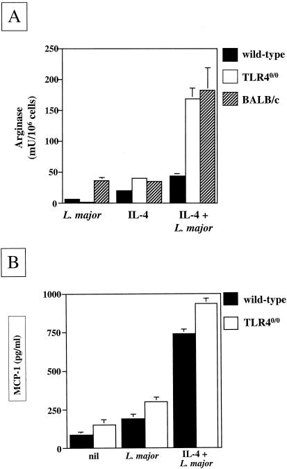
The essential role of Toll-like receptors (TLR) in innate immune responses to bacterial pathogens is increasingly recognized, but very little is known about the role of TLRs in host defense against infections with eukaryotic pathogens. For the present study, we investigated whether TLRs contribute to the innate and acquired immune response to infection with the intracellular protozoan parasite Leishmania major. Our results show that TLR4 contributes to the control of parasite growth in both phases of the immune response. We also addressed the mechanism that results in killing or growth of the intracellular parasites. Control of parasite replication correlates with the early induction of inducible nitric oxide synthase in TLR4-competent mice, whereas increased parasite survival in host cells from TLR4-deficient mice correlates with a higher activity of arginase, an enzyme known to promote parasite growth. This is the first study showing that TLR4 contributes to the effective control of Leishmania infection in vivo.


Figures

FIG. 1.
FIG. 1.
Parasite loads and protein expression in mice. (A) Parasite loads in the footpads of TLR4-competent and TLR4-deficient mice 24 h after L. major infection. Groups of wild-type (n = 4; filled circles) and TLR4-deficient (n = 4; open circles) mice were infected with 2 ×ばつ 106 L. major promastigotes. At 1 day postinfection, the numbers of viable parasites in the infected footpads were determined. Data show the results of one representative experiment of three independent experiments. Each symbol represents one mouse, and the horizontal black bars represent the means for four individual mice. *, P < 0.05. (B) Expression of iNOS, arginase, and TLR4 mRNAs in the foot pads of L. major-infected TLR4-competent and TLR4-deficient mice. The footpads of wild-type (+/+) and TLR4-deficient (0/0) mice were infected with 2 ×ばつ 106 L. major parasites. After 24 h, gene expression was analyzed in the footpads of naïve and infected mice by semiquantitative RT-PCR. Samples were standardized by densitometric comparison of the amplification of the housekeeping gene β-actin.
FIG. 2.
FIG. 2.
Parasite loads and lesion sizes during infection. (A) Parasite loads in the footpads of TLR4-competent and TLR4-deficient mice during the course of L. major infection. Groups of wild-type (filled circles) and TLR40/0 (open circles) mice were infected with 2 ×ばつ 106 L. major promastigotes. At 5, 28, and 78 days postinfection, the numbers of viable parasites in the infected footpads were determined by a limiting dilution assay. Each symbol represents one mouse, and the horizontal black bars represent the means for four individual mice. *, P < 0.05. N.D., not detectable. (B) Lesion sizes in TLR4-competent and TLR4-deficient mice during L. major infection. Groups of wild-type (n = 4; filled circles) and TLR40/0 (n = 4; open circles) mice were infected with 2 ×ばつ 106 L. major promastigotes. Values represent the lesion sizes of individual mice at the indicated times after infection. Each symbol represents one mouse, and the horizontal black bars represent the means for four individual mice. *, P < 0.05.
FIG. 2.
FIG. 2.
Parasite loads and lesion sizes during infection. (A) Parasite loads in the footpads of TLR4-competent and TLR4-deficient mice during the course of L. major infection. Groups of wild-type (filled circles) and TLR40/0 (open circles) mice were infected with 2 ×ばつ 106 L. major promastigotes. At 5, 28, and 78 days postinfection, the numbers of viable parasites in the infected footpads were determined by a limiting dilution assay. Each symbol represents one mouse, and the horizontal black bars represent the means for four individual mice. *, P < 0.05. N.D., not detectable. (B) Lesion sizes in TLR4-competent and TLR4-deficient mice during L. major infection. Groups of wild-type (n = 4; filled circles) and TLR40/0 (n = 4; open circles) mice were infected with 2 ×ばつ 106 L. major promastigotes. Values represent the lesion sizes of individual mice at the indicated times after infection. Each symbol represents one mouse, and the horizontal black bars represent the means for four individual mice. *, P < 0.05.
FIG. 3.
FIG. 3.
Cytokine and chemokine production by lymph node cells from L. major-infected wild-type and TLR40/0 mice. Groups of wild-type (filled circles) and TLR40/0 (open circles) mice were infected with 2 ×ばつ 106 L. major promastigotes in one hind footpad. At 4 weeks postinfection, 5 ×ばつ 106 popliteal lymph node cells were restimulated with 106 L. major parasites. Supernatants were harvested after 48 h and tested for their cytokine contents by ELISA. Each symbol represents one mouse, and the horizontal black bars represent the means for four individual mice. **, P > 0.05.
FIG. 4.
FIG. 4.
Parasite survival in macrophages from TLR40/0 and wild-type mice. BMMφ (5 ×ばつ 105 ml−1) from naïve wild-type (black bars) and TLR40/0 (open bars) mice were infected with 25 ×ばつ 105 L. major parasites ml−1 in the presence of IFN-γ (20 U ml−1) and TNF-α (200 U ml−1) (Th1 conditions) or IL-4 (20 U ml−1) (Th2 conditions). After 48 h of incubation, macrophages were lysed and a limiting dilution assay was performed to determine the number of viable parasites. Data show the results of one representative experiment of four independent experiments. In addition, gene expression was analyzed by semiquantitative RT-PCR. Samples were standardized by densitometric comparison with the amplification of the housekeeping gene β-actin. Data show the results of one representative experiment of two independent experiments.
FIG. 5.
FIG. 5.
(A) Differential induction of arginase activity in L. major-infected macrophages from TLR4-competent or TLR4-deficient mice. BMMφ (5 ×ばつ 105 ml−1) from naïve wild-type (black bars), TLR40/0 (white bars), and BALB/c (hatched bars) mice were cultured in the presence and/or absence of 25 ×ばつ 105 L. major parasites ml−1 and IL-4 (20 U ml−1). After 48 h, the arginase activity in macrophage lysates was measured. Data show the results of one representative experiment of five independent experiments. (B) MCP production by L. major-infected macrophages from TLR4-competent or TLR4-deficient mice. BMMφ (5 ×ばつ 105 ml−1) from naïve wild-type (black bars) and TLR40/0 (white bars) mice were cultured in the presence or absence of 25 ×ばつ 105 L. major parasites ml−1 and IL-4 (20 U ml−1). Supernatants were harvested after 48 h and tested for their MCP-1 contents by ELISA. Data show the results of one representative experiment of three independent experiments.

References

    1. Abdallahi, O. M. S., H. Bensalem, R. Augier, M. Diagana, M. De Regi, and B. Gharib. 2001. Arginase expression in peritoneal macrophages and increase in circulating polyamine levels in mice infected with Schistosoma mansoni. Cell. Mol. Life Sci. 58:1350-1357. - PMC - PubMed
    1. Aliprantis, A. O., R.-B. Yang, M. R. Mark, S. Suggett, B. Devaux, J. D. Radolf, G. R. Klimpel, P. Godowski, and A. Zychlinsky. 1999. Cell activation and apoptosis by bacterial lipoproteins through Toll-like receptor-2. Science 285:736-739. - PubMed
    1. Belkaid, Y., K. F. Hoffmann, S. Mendez, S. Kamhawi, M. C. Udey, T. A. Wynn, and D. L. Sacks. 2001. The role of interleukin (IL)-10 in the persistence of Leishmania major in the skin after healing and the therapeutic potential of anti-IL-10 receptor antibody for sterile cure. J. Exp. Med. 194:1497-1506. - PMC - PubMed
    1. Bogdan, C. 2000. The function of type I interferons in antimicrobial immunity. Curr. Opin. Immunol. 12:419-424. - PubMed
    1. Bogdan, C. 2001. Nitric oxide and the immune response. Nat. Immunol. 2:907-916. - PubMed

Publication types

MeSH terms

LinkOut - more resources

Cite

AltStyle によって変換されたページ (->オリジナル) /