This site needs JavaScript to work properly. Please enable it to take advantage of the complete set of features!
Skip to main page content
U.S. flag

An official website of the United States government

Dot gov

The .gov means it’s official.
Federal government websites often end in .gov or .mil. Before sharing sensitive information, make sure you’re on a federal government site.

Https

The site is secure.
The https:// ensures that you are connecting to the official website and that any information you provide is encrypted and transmitted securely.

NIH NLM Logo
Log in
Access keys NCBI Homepage MyNCBI Homepage Main Content Main Navigation
. 2003 Aug 18;198(4):545-56.
doi: 10.1084/jem.20030088.

Brucella evades macrophage killing via VirB-dependent sustained interactions with the endoplasmic reticulum

Affiliations

Brucella evades macrophage killing via VirB-dependent sustained interactions with the endoplasmic reticulum

Jean Celli et al. J Exp Med. .

Abstract

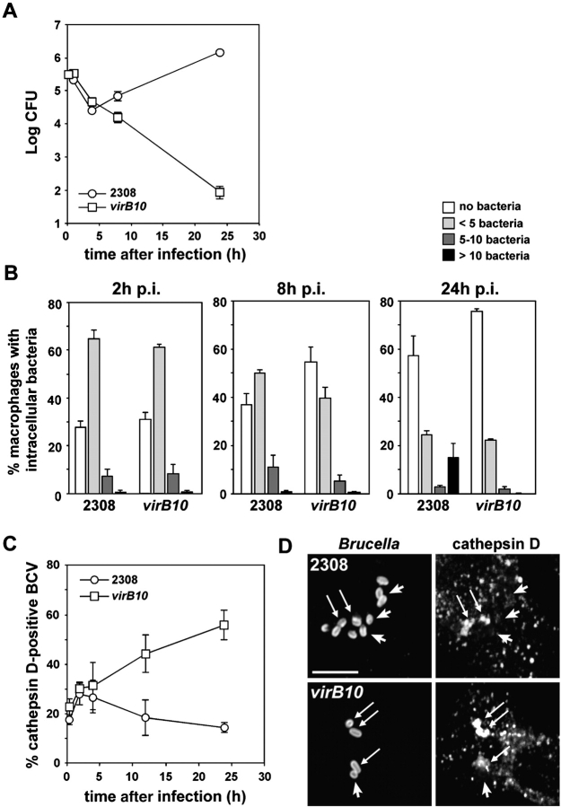
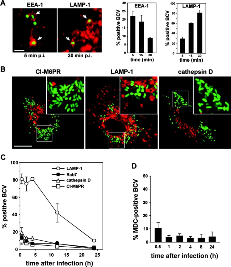
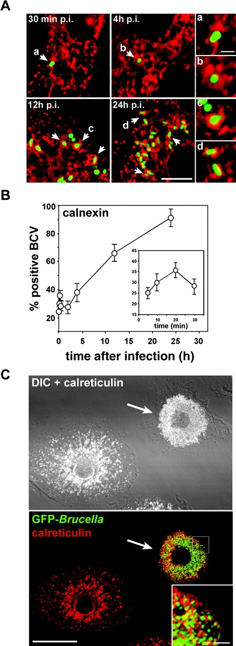
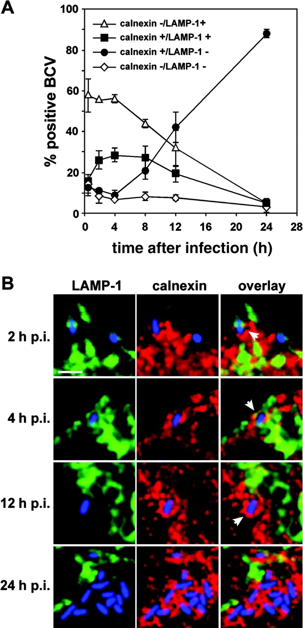
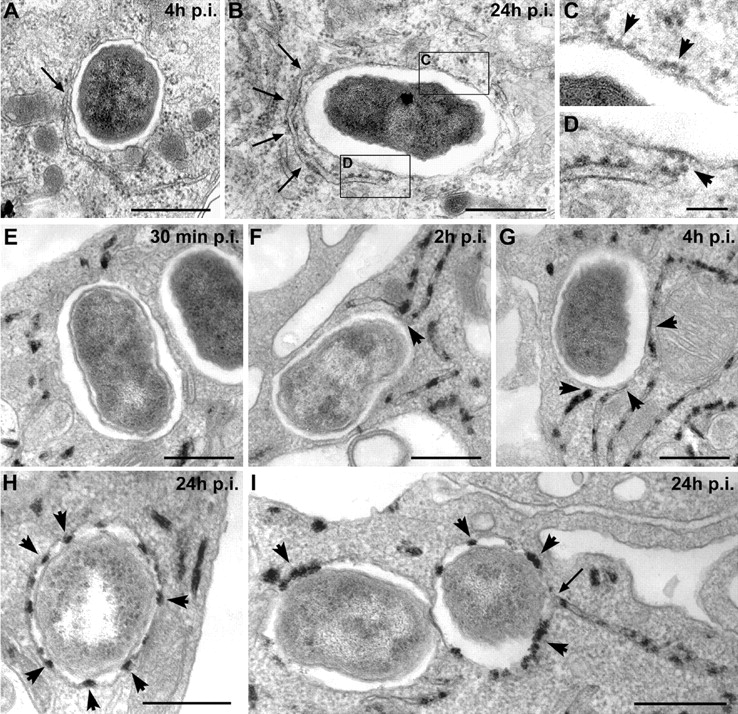
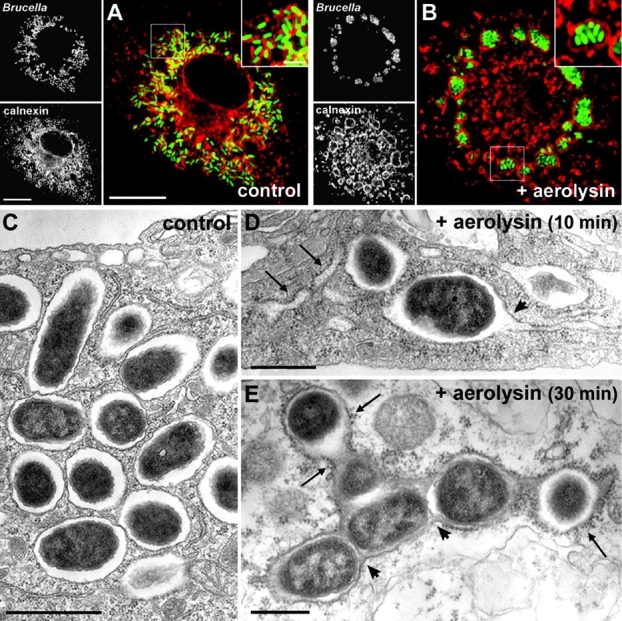
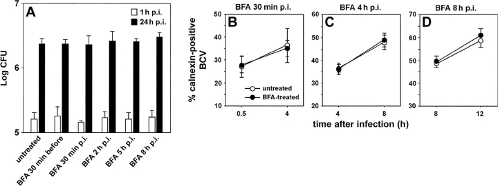
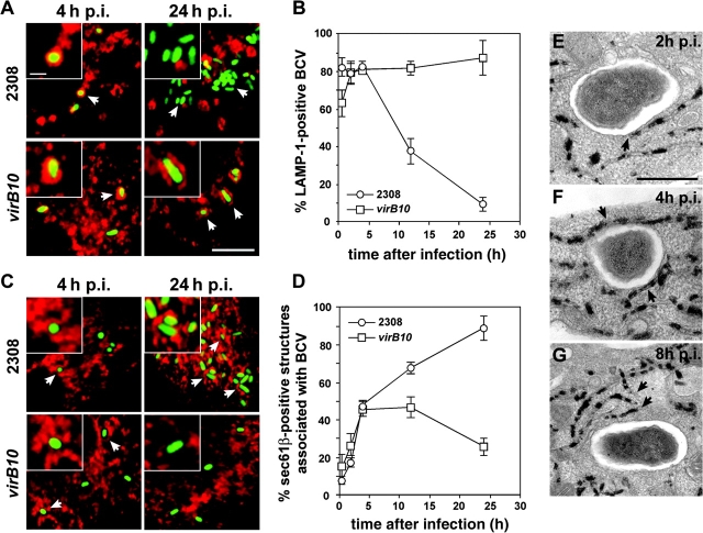
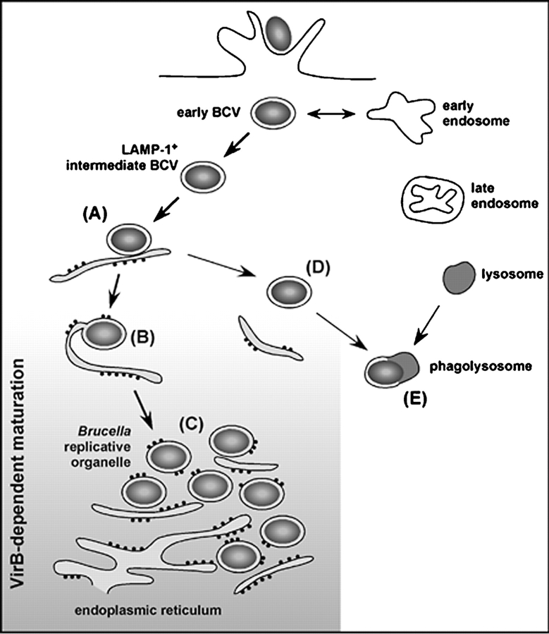
The intracellular pathogen Brucella is the causative agent of brucellosis, a worldwide zoonosis that affects mammals, including humans. Essential to Brucella virulence is its ability to survive and replicate inside host macrophages, yet the underlying mechanisms and the nature of the replicative compartment remain unclear. Here we show in a model of Brucella abortus infection of murine bone marrow-derived macrophages that a fraction of the bacteria that survive an initial macrophage killing proceed to replicate in a compartment segregated from the endocytic pathway. The maturation of the Brucella-containing vacuole involves sustained interactions and fusion with the endoplasmic reticulum (ER), which creates a replicative compartment with ER-like properties. The acquisition of ER membranes by replicating Brucella is independent of ER-Golgi COPI-dependent vesicular transport. A mutant of the VirB type IV secretion system, which is necessary for intracellular survival, was unable to sustain interactions and fuse with the ER, and was killed via eventual fusion with lysosomes. Thus, we demonstrate that live intracellular Brucella evade macrophage killing through VirB-dependent sustained interactions with the ER. Moreover, we assign an intracellular function to the VirB system, as being required for late maturation events necessary for the biogenesis of an ER-derived replicative organelle.


Figures

Figure 1.
Figure 1.
Survival and replication of Brucella strains within BMDM. BMDM were infected with either the wild-type 2308 or the virB10 mutant Brucella strains for various times. CFUs were enumerated after lysis (A) or samples were fixed and processed for immunofluorescence (B–D). (A) Representative growth curves of wild-type 2308 (しろまる) and virB10 (しろいしかく) strains inside BMDM. (B) Quantitation of 2308- or virB10-infected BMDM containing either no bacteria (open bars), less than five bacteria (light gray bars), between 5 and 10 bacteria (dark gray bars), or >10 bacteria (solid bars) at 2, 8, and 24 h after infection. Data are means ± SD of three independent experiments. (C) Quantitation of cathepsin D acquisition by BCVs. Data are means ± SD of three independent experiments. (D) Representative confocal images showing colocalization of Brucella with cathepsin D in BMDM infected with either the wild-type 2308 or the virB10 mutant Brucella strains for 24 h. Arrows indicate colocalization and arrowheads show cathepsin D BCVs. Bar, 5 μm.
Figure 2.
Figure 2.
Live Brucella interact with early but not late endocytic compartments. BMDM were infected with the GFP-expressing Brucella wild-type strain GFP-2308 for various times. Samples were fixed and processed for immunofluorescence. (A) Confocal images and quantitation of EEA-1 or LAMP-1 acquisition by early BCVs. Arrowheads show intracellular Brucella within either EEA-1+ or LAMP-1+ vacuoles. Data are means ± SD of three independent experiments. (B) Confocal images of GFP-2308–infected BMDM at 24 h after infection. Replicating Brucella do not colocalize with either CI-M6PR, LAMP-1, or cathepsin D. (C) Quantitation of LAMP-1, Rab7, CI-M6PR, and cathepsin D acquisition by BCVs during maturation. Data are means ± SD of three to five independent experiments. (D) Quantitation of MDC accumulation within BCVs. Data are means ± SD of three independent experiments. Bars, 2 μm (A) and 10 μm (B).
Figure 3.
Figure 3.
BCVs acquire ER markers during maturation. BMDM were infected with the GFP-expressing Brucella wild-type strain GFP-2308 for various times. Samples were fixed and processed for immunofluorescence. (A) Representative confocal images of a time course of GFP–Brucella interaction with ER structures. Arrowheads indicate calnexin+ BCVs. (B) Quantitation of BCV association with, and acquisition of, calnexin+ structures. Data are means ± SD of five independent experiments. (C) Overimposed confocal and differential interference contrast (DIC) images of BMDM infected with GFP Brucella for 48 h. The ER was labeled using rabbit anti–calreticulin, followed by Alexa Fluor® 594–conjugated goat anti–rabbit antibodies. The arrow indicates a Brucella-infected BMDM, whose ER is reorganized into or around BCVs. Bars, 5 μm (A), 1 μm (A insets), 20 μm (C), and 2 μm (C inset).
Figure 4.
Figure 4.
Some BCVs transiently harbor both LAMP-1 and calnexin. BMDM were infected with the wild-type GFP Brucella for various times. Samples were fixed and processed for immunofluorescence. Calnexin was detected using rabbit anti–calnexin, followed by Alexa Fluor® 594–conjugated goat anti–rabbit antibodies. LAMP-1 was detected using a rat anti–mouse LAMP-1 monoclonal antibody, followed by cyanin-5–conjugated donkey anti–rat antibodies. (A) Quantitation of calnexin/LAMP-1+ (▵), calnexin+/LAMP-1+ (▪), calnexin+/LAMP-1 (•), and calnexin/LAMP-1 (⋄) BCVs during a 24-h time course. BCVs were scored for LAMP-1 and calnexin presence from single 0.2-μm section images obtained by confocal microscopy. (B) Details from single confocal image sections showing representative BCVs at various times after infection. For clarity, the images have been artificially recolored to show bacteria in blue, LAMP-1 in green, and calnexin in red. Early BCVs (2 and 4 h after infection) harbor both markers (arrowheads) whereas late (12 h after infection) and replicative BCVs (24 h after infection) are only stained with calnexin (arrowhead). Bar, 2 μm.
Figure 5.
Figure 5.
BCVs acquire ER membranes through limited fusion with the ER. BMDM were infected with the Brucella wild-type strain 2308 for various times. Samples were processed for conventional EM (A–D) or EM cytochemistry for G6Pase detection (E–I). (A) BCVs at 4 h after infection showing a close contact with the ER (arrow). (B) BCVs at 24 h after infection surrounded by ER (arrows). (C) Enlarged view of the vacuolar membrane from B showing ribosomes studding the membrane (arrowheads). (D) Enlarged view of the contact site between ER and the vacuolar membrane showing ribosomes on the vacuolar membrane (arrowhead). (E) BCVs at 30 min after infection showing no interaction with G6Pase+ ER. (F) BCVs at 2 h after infection in close contact with G6Pase+ ER (arrowheads). (G) BCVs at 4 h after infection in intimate contact with several G6Pase+ ER compartments (arrowheads). (H) Representative G6Pase+ late BCVs at 24 h after infection (arrowheads show the intravacuolar G6Pase reaction product). (I) G6Pase+ late BCVs at 24 h after infection. The right-hand side BCV is fusing with the ER (arrow). Bars, 1 μm (A and B), 0.2 μm (C and D), and 0.5 μm (E–I).
Figure 6.
Figure 6.
Late BCVs harbor ER properties. BMDM were infected with the wild-type GFP Brucella for 36 h, and then treated with 0.5 nM proaerolysin or mock treated for 10 or 30 min before processing for either immunofluorescence (A and B) or EM (C–E). (A) Confocal images of a control untreated macrophage showing replicating GFP Brucella scattered around the nucleus in calnexin+ vacuoles (red). The inset shows individual bacteria within calnexin+ vacuoles. (B) Confocal images of an aerolysin-treated macrophage showing a dramatic relocation of GFP Brucella into giant calnexin+ vacuoles. The inset shows multiple bacteria enclosed within a large calnexin+ vacuole. (C) EM picture of an untreated macrophage showing replicating Brucella in individual vacuoles. (D) EM picture of an infected macrophage treated with proaerolysin for 10 min. Typical aerolysin-induced swelling of the ER is visible (arrows) and BCVs start to fuse with the ER (arrowhead). (E) EM picture of an infected macrophage treated with proaerolysin for 30 min. BCVs have fused together (arrowheads) into a large compartment covered with ribosomes (arrows). Bars, 10 μm (A and B), 2 μm (insets in A and B), 1 μm (C), and 0.5 μm (D and E).
Figure 7.
Figure 7.
Brucella intracellular replication does not require ER-Golgi COPI-dependent vesicular transport. (A) BMDM infected with the wild-type Brucella strain 2308 were left untreated or treated with 10 μg/ml BFA either 30 min before infection, or 30 min, 2, 5, or 8 h after infection, over a 3-h period. For each treatment, CFUs were enumerated after 1 and 24 h after infection. Data are from a representative experiment (n = 3 independent experiments). (B–D) BMDM infected with the wild-type Brucella strain GFP-2308 were left untreated or treated with 10 μg/ml BFA either from 30 min to 4 h after infection (B), 4–8 h after infection (C), or 8–12 h after infection (D). Samples were processed for immunofluorescence staining and calnexin+ BCVs were scored. Data are means ± SD of three independent experiments.
Figure 8.
Figure 8.
VirB mutant–containing vacuoles fail to sustain fusion-proficient interactions with the ER. BMDM were infected with either the wild-type Brucella strain GFP-2308 or the mutant strain GFP-virB10 for various times. Samples were processed for immunofluorescence (A–D) or EM staining for G6Pase (E–G). (A) Confocal images of infected BMDM showing acquisition of LAMP-1 on 2308 or virB10-containing vacuoles after 4 or 24 h of infection. (B) Quantitation of LAMP-1 acquisition by 2308 (しろまる)- or virB10 (しろいしかく)-containing vacuoles. Data are means ± SD of three independent experiments. (C) Confocal images of GFP Brucella–infected BMDM showing association with, or acquisition of, Sec61β+ structures on 2308 or virB10-containing vacuoles after 4 or 24 h of infection. (D) Quantitation of association of Sec61β1 structures with 2308 (しろまる)- or virB10 (しろいしかく)-containing vacuoles. Data are means ± SD of five independent experiments. (E) Staining for G6Pase in virB10-infected BMDM at 2 h after infection. The arrowhead shows a close association of BCVs with ER. (F) Staining for G6Pase in virB10-infected BMDM at 4 h after infection. The BCV is surrounded by ER (arrowheads). (G) Staining for G6Pase in virB10-infected BMDM at 8 h after infection Arrowheads show that the ER is no longer in the close vicinity of the BCV. G6Pase reaction product is not detectable inside the BCV. Bars, 5 μm (A and C), 1 μm (insets in A and C), and 0.5 μm (E–G).
Figure 9.
Figure 9.
Model for Brucella evasion of macrophage killing. After entry, intracellular Brucella resides within a vacuole (BCV) that interacts with early endosomes. These early BCVs avoid further interactions with the endocytic pathway, yet acquire LAMP-1 and are found surrounded by, or in close contact with, the ER within the first hours after infection (A). Such interactions are sustained over time and lead to limited fusion events (B), ultimately generating an ER-derived organelle permissive for Brucella replication (C). Vacuoles containing a virB-defective mutant fail to sustain fusion-proficient interactions with the ER (D) and ultimately fuse with lysosomes (E). The shaded area designates the VirB-dependent maturation events of BCVs described in this work. ER membrane acquisition by BCVs is illustrated by the localized presence of ribosomes.

References

    1. Knodler, L.A., J. Celli, and B.B. Finlay. 2001. Pathogenic trickery: deception of host cell processes. Nat. Rev. Mol. Cell Biol. 2:578–588. - PubMed
    1. Moreno, E., E. Stackebrandt, M. Dorsch, J. Wolters, M. Busch, and H. Mayer. 1990. Brucella abortus 16S rRNA and lipid A reveal a phylogenetic relationship with members of the alpha-2 subdivision of the class Proteobacteria. J. Bacteriol. 172:3569–3576. - PMC - PubMed
    1. Moreno, E., and I. Moriyon. 2001. The genus Brucella. The Prokaryotes. M. Dorkin, S. Falkow, E. Rosenberg, K.-H. Schleifer, and E. Stackebrandt, editors. Springer-Verlag, New York.
    1. Gorvel, J.P., and E. Moreno. 2002. Brucella intracellular life: from invasion to intracellular replication. Vet. Microbiol. 90:281–297. - PubMed
    1. Hong, P.C., R.M. Tsolis, and T.A. Ficht. 2000. Identification of genes required for chronic persistence of Brucella abortus in mice. Infect. Immun. 68:4102–4107. - PMC - PubMed

Publication types

MeSH terms

Cite

AltStyle によって変換されたページ (->オリジナル) /