This site needs JavaScript to work properly. Please enable it to take advantage of the complete set of features!
Skip to main page content
U.S. flag

An official website of the United States government

Dot gov

The .gov means it’s official.
Federal government websites often end in .gov or .mil. Before sharing sensitive information, make sure you’re on a federal government site.

Https

The site is secure.
The https:// ensures that you are connecting to the official website and that any information you provide is encrypted and transmitted securely.

NIH NLM Logo
Log in
Access keys NCBI Homepage MyNCBI Homepage Main Content Main Navigation
. 2002 Dec;76(24):12463-72.
doi: 10.1128/jvi.76.24.12463-12472.2002.

Covalent modifications of the ebola virus glycoprotein

Affiliations

Covalent modifications of the ebola virus glycoprotein

Scott A Jeffers et al. J Virol. 2002 Dec.

Abstract

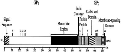
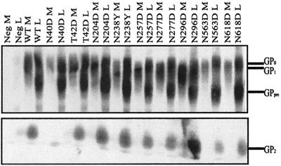
The role of covalent modifications of the Ebola virus glycoprotein (GP) and the significance of the sequence identity between filovirus and avian retrovirus GPs were investigated through biochemical and functional analyses of mutant GPs. The expression and processing of mutant GPs with altered N-linked glycosylation, substitutions for conserved cysteine residues, or a deletion in the region of O-linked glycosylation were analyzed, and virus entry capacities were assayed through the use of pseudotyped retroviruses. Cys-53 was the only GP(1) ( approximately 130 kDa) cysteine residue whose replacement resulted in the efficient secretion of GP(1), and it is therefore proposed that it participates in the formation of the only disulfide bond linking GP(1) to GP(2) ( approximately 24 kDa). We propose a complete cystine bridge map for the filovirus GPs based upon our analysis of mutant Ebola virus GPs. The effect of replacement of the conserved cysteines in the membrane-spanning region of GP(2) was found to depend on the nature of the substitution. Mutations in conserved N-linked glycosylation sites proved generally, with a few exceptions, innocuous. Deletion of the O-linked glycosylation region increased GP processing, incorporation into retrovirus particles, and viral transduction. Our data support a common evolutionary origin for the GPs of Ebola virus and avian retroviruses and have implications for gene transfer mediated by Ebola virus GP-pseudotyped retroviruses.


Figures

FIG. 1.
FIG. 1.
Schematic representation of Ebola virus GP. The GP1 and GP2 subunits of GP are drawn to scale (residue numbers are indicated below the diagram). The positions of the signal sequence (cross-hatching), conserved cysteine residues (S), the mucin-like region (region of O-linked glycosylation; black), the furin cleavage site, the fusion peptide (vertical lines), the coiled-coil domain (diagonal lines), and the membrane-spanning domain (horizontal line) are indicated.
FIG. 2.
FIG. 2.
RIPA of GP in 293 cells. The GPs listed in Table 1 were expressed in 293 cells by using a VV-T7 system and were radiolabeled with [35S]cysteine. They were then immunoprecipitated with a rabbit anti-Ebola virus SGP-GP serum, the reduced proteins were analyzed by SDS-10% PAGE under reducing conditions, and autoradiography was performed. Immunoprecipitated GPs secreted or released into the medium (M) or associated with the cell monolayer (L) were run side by side; only the relevant portions of the gels are shown. Detection of GP2 is shown only in monolayer lanes. The migration positions of GP1, GP2, and GPpre are indicated on the left; GPpre is an uncleaved immature or precursor form of GP that is primarily associated with the ER (27). Asterisks in the GP1 region identify increased levels of this GP in the medium relative to cell-associated GP1, compared to the levels in the wild type (WT). Asterisks in the GP2 region identify faster-migrating forms of GP2. There are cross-reactive species migrating just slower and somewhat faster than wild-type GP2. Neg, transfection of pTM1(ΔNcoI) vector into 293 cells infected with a recombinant vaccinia virus (vTF7-3) expressing T7 RNA polymerase.
FIG. 3.
FIG. 3.
Migration of GP under nonreducing conditions. Shown is an autoradiogram of SDS-PAGE analysis (under nonreducing conditions) of wild-type (WT) GP and of the proteins analyzed in Fig. 2 that showed increased release of GP1 into the medium. Immunoprecipitated GPs secreted or released into the medium (M) or associated with the cell monolayer (L) were run side by side. The migration positions of GP1, GP2, and GPpre are indicated on the left.
FIG. 4.
FIG. 4.
Analysis of the expression and incorporation into pseudotyped retroviruses of Ebola virus GPs with substitutions of ectodomain cysteine residues. φNX cells were transfected with plasmids encoding Ebola virus GPs. The cell lysates (L) and viral particles collected from the culture medium (M) were analyzed by SDS-PAGE (8.5% acrylamide) and immunoblotting with anti-Ebola virus SGP-GP antibody. Analysis of a cell lysate aliquot that was treated with PNGase F (+), which removes N-linked glycosylation, is also shown. The migration positions of mature GP1, GP0 (the glycosylated but uncleaved form), GPpre (the N-glycosylated but not O-glycosylated uncleaved form), and deglycosylated GP0 and GPpre are indicated. WT, wild type; Neg, ΦNX cells transfected with pcDNA3 vector.
FIG. 5.
FIG. 5.
Analysis of the expression and incorporation into pseudotyped retroviruses of Ebola virus GPs with substitutions of membrane-spanning domain cysteine residues. Analysis was conducted as described in the legend to Fig. 4. The migration positions of mature GP1, GP0 (the glycosylated but uncleaved form), GPpre (the N-glycosylated but not O-glycosylated uncleaved form), and deglycosylated GP0 and GPpre are indicated. WT, wild type; Neg, ΦNX cells transfected with pcDNA3 vector.
FIG. 6.
FIG. 6.
Analysis of the expression and incorporation into pseudotyped retroviruses of Ebola virus GPs with substitutions eliminating sites of N-linked glycosylation. Analysis was conducted as described in the legend to Fig. 4, except that no PNGase F was used. The migration positions of mature GP1, GP0 (the glycosylated but uncleaved form), GPpre (the N-glycosylated but not O-glycosylated uncleaved form), and GP2 are indicated. WT, wild type; Neg, ΦNX cells transfected with pcDNA3 vector.
FIG. 7.
FIG. 7.
Analysis of the expression and incorporation into pseudotyped retroviruses of the Δ309-489 Ebola virus GP. Analysis was conducted as described in the legend to Fig. 4. The migration positions of the mature GP1, GP0 (the glycosylated but uncleaved form), GPpre (the N-glycosylated but not O-glycosylated uncleaved form), and deglycosylated GP0 and GPpre forms of wild-type (WT) GP and of the GP1, GP0, and deglycosylated GP1 and GP0 forms of Δ309-489 GP are indicated. Neg, ΦNX cells transfected with pcDNA3 vector.
FIG. 8.
FIG. 8.
Analysis of the glycosylation of the Δ309-489 Ebola virus GP incorporated into pseudotyped retroviruses. Analysis was conducted as described in the legend to Fig. 4, except that aliquots of the samples were treated with PNGase F, with a combination of PNGase F, sialidase A, and endo-O-glycosidase, or with a combination of the previous three enzymes and β(1-4)-galactosidase and glucosaminidase. The migration positions of the mature GP1 forms of the wild-type (WT) and Δ309-489 GPs are indicated. In this experiment, a glycosylated serum protein showing a mobility intermediate between those of wild-type GP1 and Δ309-489 GP1 was detected. The heterogeneous mobility of the PNGase F-treated proteins was indicative of the incomplete removal of N glycosylation. Neg, ΦNX cells transfected with pcDNA3 vector.
FIG. 9.
FIG. 9.
Cystine bridge model for Ebola virus GP and comparison of GP2 to the Rous sarcoma virus GP TM subunit. Representational elements for the mucin-like region, the fusion peptides, the coiled-coil domains, and the membrane-spanning domains are identical to those used in Fig. 1. (A) The cystine bridge arrangement in the Ebola virus GP deduced from the results presented here and elsewhere (10, 16, 36) and the critical N-glycosylation sites (Y) discussed in the text are depicted. (B) A proposed cystine bridge model for the TM protein of Rous sarcoma virus (an ASLV) (3, 10) is presented for comparison with that for Ebola virus GP2.

References

    1. Chan, S. Y., R. F. Speck, M. C. Ma, and M. A. Goldsmith. 2000. Distinct mechanisms of entry by envelope glycoproteins of Marburg and Ebola (Zaire) viruses. J. Virol. 74:4933-4937. - PMC - PubMed
    1. Chepurnov, A. A., M. N. Tuzova, V. A. Ternovoy, and I. V. Chernukhin. 1999. Suppressive effect of Ebola virus on T cell proliferation in vitro is provided by a 125-kDa GP viral protein. Immunol. Lett. 68:257-261. - PubMed
    1. Delos, S. E., and J. M. White. 2000. Critical role for the cysteines flanking the internal fusion peptide of avian sarcoma/leukosis virus envelope glycoprotein. J. Virol. 74:9738-9741. - PMC - PubMed
    1. Ellgaard, L., M. Molinari, and A. Helenius. 1999. Setting the standards: quality control in the secretory pathway. Science 286:1882-1888. - PubMed
    1. Elroy-Stein, O., T. R. Fuerst, and B. Moss. 1989. Cap-independent translation of mRNA conferred by encephalomyocarditis virus 5′ sequence improves the performance of the vaccinia virus/bacteriophage T7 hybrid expression system. Proc. Natl. Acad. Sci. USA 86:6126-6130. - PMC - PubMed

Publication types

Substances

LinkOut - more resources

Cite

AltStyle によって変換されたページ (->オリジナル) /