This site needs JavaScript to work properly. Please enable it to take advantage of the complete set of features!
Skip to main page content
U.S. flag

An official website of the United States government

Dot gov

The .gov means it’s official.
Federal government websites often end in .gov or .mil. Before sharing sensitive information, make sure you’re on a federal government site.

Https

The site is secure.
The https:// ensures that you are connecting to the official website and that any information you provide is encrypted and transmitted securely.

NIH NLM Logo
Log in
Access keys NCBI Homepage MyNCBI Homepage Main Content Main Navigation
. 2001 Feb;75(4):2010-3.
doi: 10.1128/JVI.75.4.2010-2013.2001.

Exposure to low pH is not required for penetration of mosquito cells by Sindbis virus

Affiliations

Exposure to low pH is not required for penetration of mosquito cells by Sindbis virus

R Hernandez et al. J Virol. 2001 Feb.

Abstract

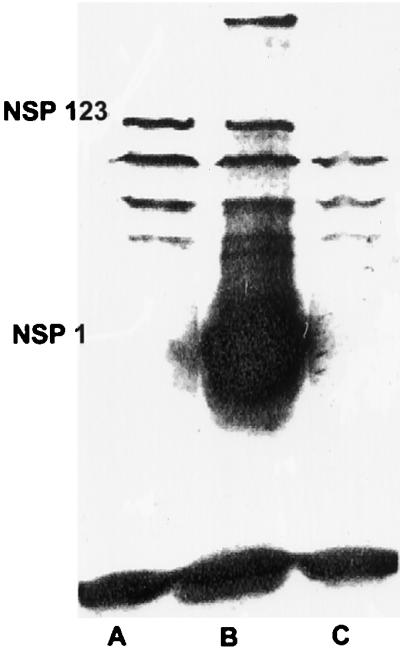
It is widely held that the penetration of cells by alphaviruses is dependent on exposure to the acid environment of an endosome. The alphavirus Sindbis virus replicates in both vertebrate and invertebrate cell cultures. We have found that exposure to an acid environment may not be required for infection of cells of the insect host. In this work, we investigated the effects of two agents (NH(4)Cl and chloroquine), which raise the pH of intracellular compartments (lysosomotropic weak bases) on the infection and replication of Sindbis virus in cells of the insect host Aedes albopictus. The results show that both of these agents increase the pH of endosomes, as indicated by protection against diphtheria toxin intoxication. NH(4)Cl blocked the production of infectious virus and blocked virus RNA synthesis when added prior to infection. Chloroquine, in contrast to its effect on vertebrate cells, had no inhibitory effect on infectious virus production in mosquito cells even when added prior to infection. Treatment with NH(4)Cl did not prevent the penetration of virus RNA into the cell cytoplasm or translation of the RNA to produce a precursor to virus nonstructural proteins. These data suggest that while these two drugs raise the pH of endosomes, they do not block insect cell penetration. These data support previous results published by our laboratory suggesting that exposure to an acid environment within the cell may not be an obligatory step in the process of infection of cells by alphaviruses.


Figures

FIG. 1
FIG. 1
Protein synthesis in diphtheria toxin-treated A. albopictus cells. Cellular protein synthesis was measured in the presence (▴) and absence (∗) of 15 mM NH4Cl (▵) or 5.0 mM chloroquine (しかく). Diphtheria toxin (List Biological Laboratories, Campbell, Calif.) was nicked with trypsin (17). Cells were treated with various concentrations of diphtheria toxin for 2 h and then labeled with Tran35S-label (cysteine-methionine; ICN, Costa Mesa, Calif.) for 1 h. Monolayers were then washed with PBS, and radioactivity incorporated into trichloroacetic acid-precipitable material was quantitated as described previously (18).
FIG. 2
FIG. 2
Production and processing of Sindbis virus nonstructural proteins in A. albopictus cells infected in the presence (lane A) and absence (lane B) of 15 mM NH4Cl. Sindbis virus-infected or mock-infected (lane C) mosquito cells were solubilized with lysis buffer as described previously (42). Proteins separated by SDS-PAGE on a 10.8% gel were transferred to a polyvinylidene difluoride membrane as described by Burnette (9). The membrane was air dried for 30 min at room temperature and then soaked in blocking solution (10% Carnation instant nonfat dry milk in PBS) with 1% goat serum for 1 h with constant, moderate agitation. The membrane was then incubated in 10% Carnation instant nonfat dry milk–0.05% Tween 20 in PBS–1% goat serum (incubation solution) containing a 1:100 dilution of rabbit anti-Sindbis virus nonstructural protein nsP1 antibody (provided by J. H. Strauss, California Institute of Technology, Pasadena) at room temperature for 1 h with constant, moderate agitation. After three 5-min washes at room temperature with 0.05% Tween 20 in PBS–1% goat serum (washing solution), the membrane was incubated for 1 h at room temperature in 100 ml of incubation solution containing 10 μCi of 125I-labeled goat anti-rabbit immunoglobulin G (DuPont New England Nuclear, Boston, Mass.). The membrane was then carefully washed four times with washing solution, air dried for 30 min at room temperature, and exposed to Kodak XAR-5 film.

References

    1. Abell B A, Brown D T. Sindbis virus membrane fusion is mediated by reduction of glycoprotein disulfide bridges at the cell surface. J Virol. 1993;67:5496–5501. - PMC - PubMed
    1. Adams R H, Brown D T. Inhibition of Sindbis virus maturation after treatment of infected cells with trypsin. J Virol. 1982;41:692–702. - PMC - PubMed
    1. Anthony R P, Brown D T. Protein-protein interactions in an alphavirus membrane. J Virol. 1991;65:1187–1194. - PMC - PubMed
    1. Anthony R P, Paredes A M, Brown D T. Disulfide bonds are essential for the stability of the Sindbis virus envelope. Virology. 1992;190:330–336. - PubMed
    1. Baric R S, Carlin L J, Johnston R E. Requirement for host transcription in the replication of Sindbis virus. J Virol. 1983;45:200–205. - PMC - PubMed

Publication types

LinkOut - more resources

Cite

AltStyle によって変換されたページ (->オリジナル) /