This site needs JavaScript to work properly. Please enable it to take advantage of the complete set of features!
Skip to main page content
U.S. flag

An official website of the United States government

Dot gov

The .gov means it’s official.
Federal government websites often end in .gov or .mil. Before sharing sensitive information, make sure you’re on a federal government site.

Https

The site is secure.
The https:// ensures that you are connecting to the official website and that any information you provide is encrypted and transmitted securely.

NIH NLM Logo
Log in
Access keys NCBI Homepage MyNCBI Homepage Main Content Main Navigation
. 2000 Jul 17;192(2):249-58.
doi: 10.1084/jem.192.2.249.

Salmonella exploits caspase-1 to colonize Peyer's patches in a murine typhoid model

Affiliations

Salmonella exploits caspase-1 to colonize Peyer's patches in a murine typhoid model

D M Monack et al. J Exp Med. .

Abstract

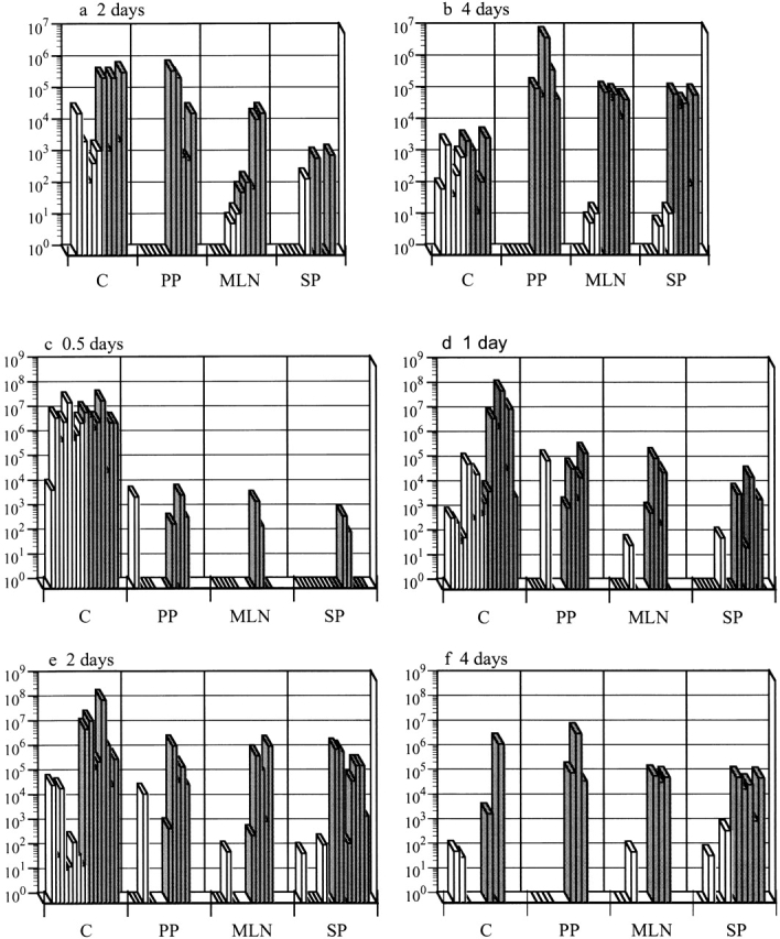
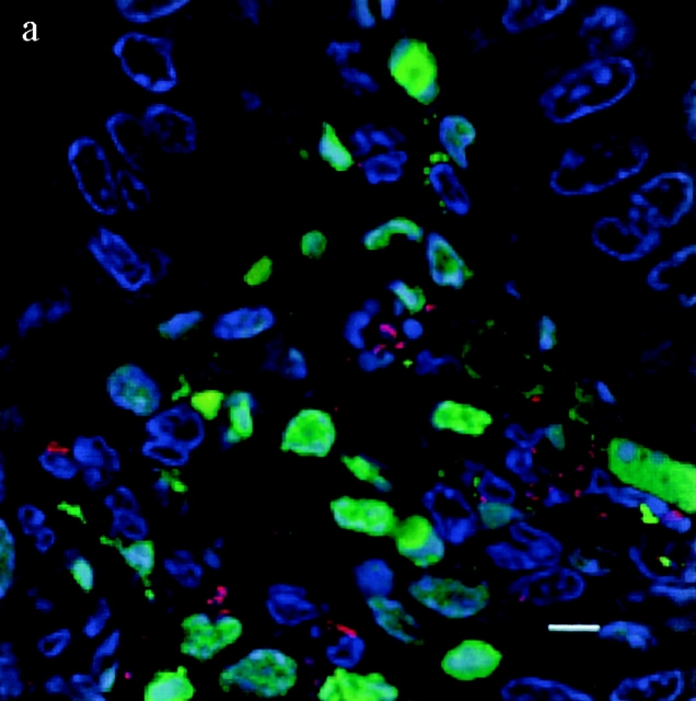
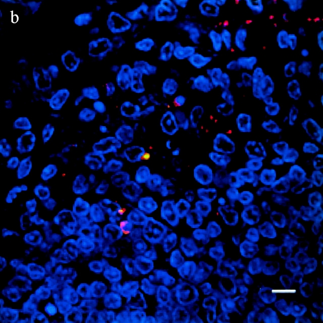
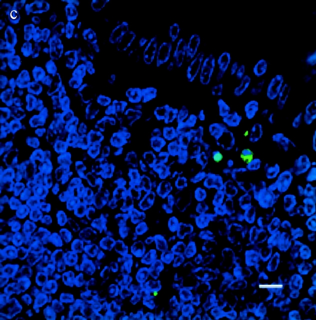
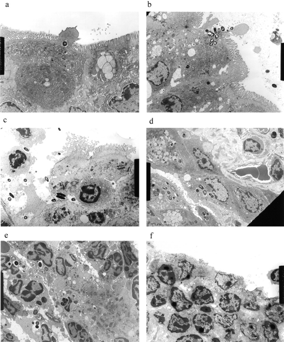
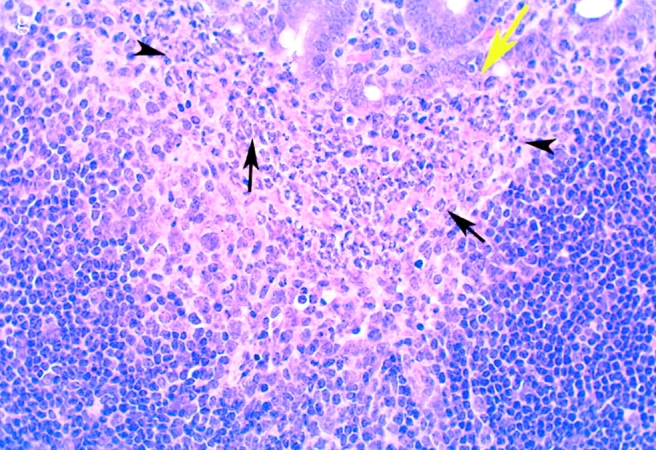
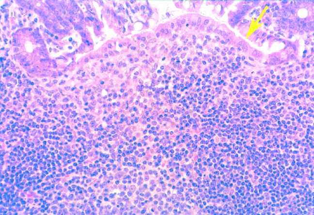
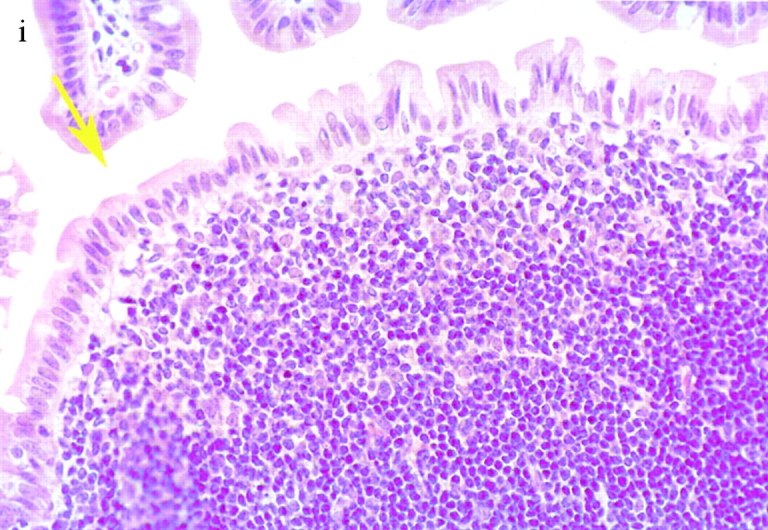
Salmonella typhimurium invades host macrophages and induces apoptosis and the release of mature proinflammatory cytokines. SipB, a protein translocated by Salmonella into the cytoplasm of macrophages, is required for activation of Caspase-1 (Casp-1, an interleukin [IL]-1beta-converting enzyme), which is a member of a family of cysteine proteases that induce apoptosis in mammalian cells. Casp-1 is unique among caspases because it also directly cleaves the proinflammatory cytokines IL-1beta and IL-18 to produce bioactive cytokines. We show here that mice lacking Casp-1 (casp-1(-/)- mice) had an oral S. typhimurium 50% lethal dose (LD(50)) that was 1,000-fold higher than that of wild-type mice. Salmonella breached the M cell barrier of casp-1(-/)- mice efficiently; however, there was a decrease in the number of apoptotic cells, intracellular bacteria, and the recruitment of polymorphonuclear lymphocytes in the Peyer's patches (PP) as compared with wild-type mice. Furthermore, Salmonella did not disseminate systemically in the majority of casp-1(-/)- mice, as demonstrated by significantly less colonization in the PP, mesenteric lymph nodes, and spleens of casp-1(-/)- mice after an oral dose of S. typhimurium that was 100-fold higher than the LD(50). The increased resistance in casp-1(-/)- animals appears specific for Salmonella infection since these mice were susceptible to colonization by another enteric pathogen, Yersinia pseudotuberculosis, which normally invades the PP. These results show that Casp-1, which is both proapoptotic and proinflammatory, is essential for S. typhimurium to efficiently colonize the cecum and PP and subsequently cause systemic typhoid-like disease in mice.


Figures

Figure 1
Figure 1
Casp-1−/− mice are resistant to S. typhimurium colonization after an oral infection. Wild-type mice (gray bars) and casp-1 / mice (white bars) were inoculated intragastrically with wild-type S. typhimurium. The tissue colonization of mice inoculated with 2 ×ばつ 107 CFU of S. typhimurium SL1344 was determined for (a) 2 and (b) 4 d after inoculation. C, cecum; PP, Peyer's patches; MLN, mesenteric lymph nodes; SP, spleen. n = 5 for both days. Tissue colonization after intragastric inoculation with 1010 CFU of S. typhimurium SL1344 for (c) 0.5, (d) 1, (e) 2, and (f) 4 d after inoculation. Cecum and spleen, n = 8 for days 0.5, 1, and 2; n = 3 for day 4. PP and MLNs, n = 5 for days 0.5, 1, and 2; n = 3 for day 4. Day 1 for PP and MLNs for wild-type compared with casp-1 −/ mice: P = 0.0236 and P = 0.0343, respectively. Day 2 for PP, MLNs, spleens, and livers: P = 0.0132, P = 0.0343, P = 0.0025, and P = .0283, respectively. Day 4 for PP, MLNs, spleens, and livers: P = 0.0369, P = 0.0038, P = 0.0207, and P = 0.0278, respectively. Rectangles on the x axis represent zero bacterial colonies present in undiluted tissue. These data represent results from one experiment, performed on age-matched casp-1 −/ and wild-type mice, which is representative of three experiments. The variability in mouse colonization at early times is reproducible and typical of what is seen after an oral inoculation.
Figure 2
Figure 2
TUNEL+ cells colocalize with S. typhimurium in the dome area of PP from casp-1 +/+ mice. Cross sections of PP from ligated intestinal loops performed in (a) wild-type or (b) casp-1 −/ mice infected with wild-type Salmonella or (c) wild-type mice inoculated with LB for 1 h were fixed and stained for apoptotic nuclei in green, Salmonella in red, and host cell nuclei in blue. The scalebar represents 8 μm in a and 10 μm in b and c. Images are oriented such that the intestinal lumen is at the top and the lymphoid follicle (not shown) is toward the bottom.
Figure 2
Figure 2
TUNEL+ cells colocalize with S. typhimurium in the dome area of PP from casp-1 +/+ mice. Cross sections of PP from ligated intestinal loops performed in (a) wild-type or (b) casp-1 −/ mice infected with wild-type Salmonella or (c) wild-type mice inoculated with LB for 1 h were fixed and stained for apoptotic nuclei in green, Salmonella in red, and host cell nuclei in blue. The scalebar represents 8 μm in a and 10 μm in b and c. Images are oriented such that the intestinal lumen is at the top and the lymphoid follicle (not shown) is toward the bottom.
Figure 3
Figure 3
(continues on facing page). S. typhimurium induces inflammation in wild-type PP but not in casp-1 −/ PP. Transmission electron micrographs of infected PP revealed bacterial invasion of M cells at 0.5 h in wild-type (a) and casp-1 −/ (b) PP, and at 1 h in (c) extracellular Salmonella in wild-type and (d) intracellular bacteria in casp-1 −/ PP. At 3 h, wild-type PP dome revealed an infiltration of PMNs and intracellular Salmonella (e), whereas casp-1 −/ PP revealed an absence of PMNs and bacteria (f). Hematoxylin and eosin stains of PP from casp-1 +/+ (g) mice infected for 3 h revealed clusters of PMNs (black arrows) and pyknotic cells (arrowheads), whereas casp-1 −/ PP did not (h). Uninfected, wild-type PP are shown for comparison (i). Yellow arrows point to the PP dome area. Serial tissue sections were stained with anti-Salmonella antibody to confirm that the PP shown was infected. All sections are oriented with intestinal lumen at the top and lymphoid follicle at the bottom.
Figure 3
Figure 3
(continues on facing page). S. typhimurium induces inflammation in wild-type PP but not in casp-1 −/ PP. Transmission electron micrographs of infected PP revealed bacterial invasion of M cells at 0.5 h in wild-type (a) and casp-1 −/ (b) PP, and at 1 h in (c) extracellular Salmonella in wild-type and (d) intracellular bacteria in casp-1 −/ PP. At 3 h, wild-type PP dome revealed an infiltration of PMNs and intracellular Salmonella (e), whereas casp-1 −/ PP revealed an absence of PMNs and bacteria (f). Hematoxylin and eosin stains of PP from casp-1 +/+ (g) mice infected for 3 h revealed clusters of PMNs (black arrows) and pyknotic cells (arrowheads), whereas casp-1 −/ PP did not (h). Uninfected, wild-type PP are shown for comparison (i). Yellow arrows point to the PP dome area. Serial tissue sections were stained with anti-Salmonella antibody to confirm that the PP shown was infected. All sections are oriented with intestinal lumen at the top and lymphoid follicle at the bottom.
Figure 4
Figure 4
S. typhimurium CFU decrease in casp-1 −/ PP. Ligated intestinal loops were performed for 0.5 and 3 h and gentamicin-protected CFU per PP was determined. Lanes 1 (n = 9) and 5 (n = 10), casp-1 +/+ mice infected with SL1344. Lanes 2 (n = 9) and 6 (n = 11), casp-1 +/+ mice infected with noninvasive mutant BJ66 3. Lanes 3 (n = 10) and 7 (n = 13,) casp-1 −/ mice infected with SL1344. Lanes 4 (n = 12) and 8 (n = 12), casp-1 −/ mice infected with BJ66. Unpaired t test revealed P < 0.0001 and P = 0.0013 for SL1344 in casp-1 −/ compared with casp-1 +/+ at 0.5 and 3 h, respectively; P = 0.0001 for SL1344 in casp-1 +/+ mice for 0.5 versus 3 h; P = 0.0028 for SL1344 in casp-1 −/ mice for 0.5 versus 3 h. The noninvasive mutant produced numbers that were not statistically different between the two mouse strains. The mean and standard deviations are shown from two separate experiments.
Figure 5
Figure 5
casp-1 −/ mice are susceptible to an i.p. route of inoculation with S. typhimurium. 100 SL1344 bacteria were injected into the peritoneal cavity of casp-1 null (▪) and wild-type (•) mice. The percentage of survival was recorded for 14 d. All casp-1–deficient mice died by day 7.
Figure 6
Figure 6
Activated peritoneal macrophages from wild-type and casp-1 −/ mice kill S. typhimurium in the absence of apoptosis. Peritoneal macrophages from wild-type mice were infected with opsonized, stationary phase SL1344 (white bars) or the noninvasive S. typhimurium mutant, BJ66 (black bars). Peritoneal macrophages from casp-1 −/ mice were infected with opsonized, stationary phase SL1344 (gray bars) or the noninvasive S. typhimurium mutant, BJ66 (hatched bars). The multiplicity of infection was 10 bacteria per macrophage. Under these conditions, there was no macrophage death as measured by assaying the culture supernatants for the presence of lactate dehydrogenase with a Cytotox96 Cell Death Kit (Promega).
Figure 7
Figure 7
casp-1 −/ mice are colonized with Y. pseudotuberculosis. Wild-type mice (gray bars) and casp-1 −/− mice (white bars) were inoculated intragastrically with a wild-type Y. pseudotuberculosis strain, YPIIIpYV. C, cecum; PP, Peyer's patches; MLN, mesenteric lymph nodes; SP, spleen. n = 3 for day 2; n = 2 for day 4.

References

    1. Darwin K.H., Miller V.L. Molecular basis of the interaction of Salmonella with the intestinal mucosa. Clin. Microbiol. Rev. 1999;12:405–428. - PMC - PubMed
    1. Jones B.D., Falkow S. Salmonellosishost immune responses and bacterial virulence determinants. Annu. Rev. Immunol. 1996;14:533–561. - PubMed
    1. Jones B.D., Falkow S. Identification and characterization of a Salmonella typhimurium oxygen-regulated gene required for bacterial internalization. Infect. Immun. 1994;62:3745–3752. - PMC - PubMed
    1. Baumler A.J., Tsolis R.M., Valentine P.J., Ficht T.A., Heffron F. Synergistic effect of mutations in invA and lpfC on the ability of Salmonella typhimurium to cause murine typhoid. Infect. Immun. 1997;65:2254–2259. - PMC - PubMed
    1. Behlau I., Miller S.I. A PhoP-repressed gene promotes Salmonella typhimurium invasion of epithelial cells. J. Bacteriol. 1993;175:4475–4484. - PMC - PubMed

Publication types

Cite

AltStyle によって変換されたページ (->オリジナル) /