Diagnosis and clinical virology of Lassa fever as evaluated by enzyme-linked immunosorbent assay, indirect fluorescent-antibody test, and virus isolation
- PMID: 10878062
- PMCID: PMC86994
- DOI: 10.1128/JCM.38.7.2670-2677.2000
Diagnosis and clinical virology of Lassa fever as evaluated by enzyme-linked immunosorbent assay, indirect fluorescent-antibody test, and virus isolation
Abstract
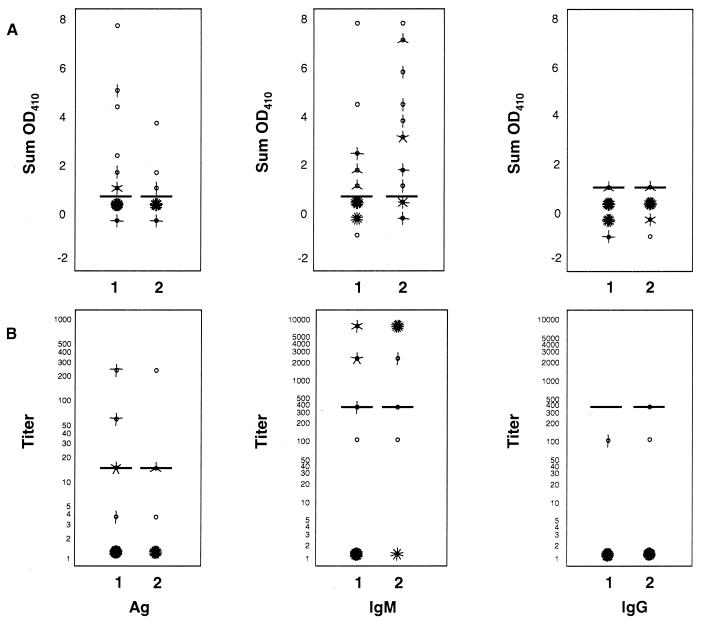
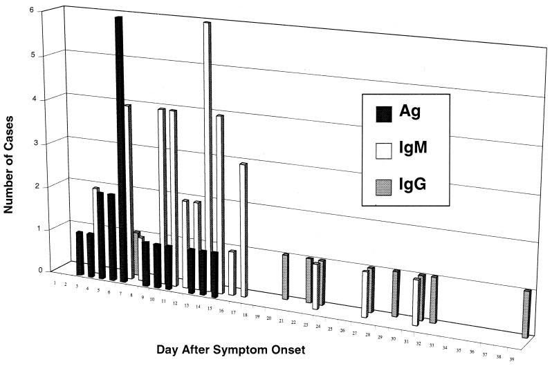
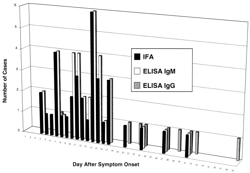
The Lassa virus (an arenavirus) is found in West Africa, where it sometimes causes a severe hemorrhagic illness called Lassa fever. Laboratory diagnosis has traditionally been by the indirect fluorescent-antibody (IFA) test. However, enzyme-linked immunosorbent assays (ELISAs) for Lassa virus antigen and immunoglobulin M (IgM) and G (IgG) antibodies have been developed that are thought to be more sensitive and specific. We compared ELISA and IFA testing on sera from 305 suspected cases of Lassa fever by using virus isolation with a positive reverse transcription-PCR (RT-PCR) test as the "gold standard." Virus isolation and RT-PCR were positive on 50 (16%) of the 305 suspected cases. Taken together, Lassa virus antigen and IgM ELISAs were 88% (95% confidence interval [CI], 77 to 95%) sensitive and 90% (95% CI, 88 to 91%) specific for acute infection. Due to the stringent gold standard used, these likely represent underestimates. Diagnosis could often be made on a single serum specimen. Antigen detection was particularly useful in providing early diagnosis as well as prognostic information. Level of antigenemia varied inversely with survival. Detection by ELISA of IgG antibody early in the course of illness helped rule out acute Lassa virus infection. The presence of IFA during both acute and convalescent stages of infection, as well as significant interobserver variation in reading the slides, made interpretation difficult. However, the assay provided useful prognostic information, the presence of IFA early in the course of illness correlating with death. The high sensitivity and specificity, capability for early diagnosis, and prognostic value of the ELISAs make them the diagnostic tests of choice for the detection of Lassa fever.
Figures
References
-
- Arnold R B, Gary G W., Jr A neutralization test survey for Lassa fever activity in Lassa, Nigeria. Trans R Soc Trop Med Hyg. 1977;71:152–154. - PubMed
-
- Bowen M D, Peters C J, Nichol S T. Phylogenetic analysis of the Arenaviridae: patterns of virus evolution and evidence for cospeciation between Arenaviruses and their rodent hosts. Mol Phylogenet Evol. 1997;8:301–316. - PubMed
-
- Buchmeier M, Welsh R, Dutko F, Oldstone M. The virology and immunobiology of lymphocytic choriomeningitis virus infection. Adv Immunol. 1980;30:275–331. - PubMed
-
- Byrne J, Ahmed R, Oldstone M. Biology of cloned cytotoxic T lymphocytes specific for lymphocytic choriomeningitis virus. I. Generation and recognition of virus strains and H-2b mutants. J Immunol. 1984;133:433–439. - PubMed
Publication types
MeSH terms
Substances
LinkOut - more resources
Full Text Sources
Other Literature Sources