This site needs JavaScript to work properly. Please enable it to take advantage of the complete set of features!
Skip to main page content
U.S. flag

An official website of the United States government

Dot gov

The .gov means it’s official.
Federal government websites often end in .gov or .mil. Before sharing sensitive information, make sure you’re on a federal government site.

Https

The site is secure.
The https:// ensures that you are connecting to the official website and that any information you provide is encrypted and transmitted securely.

NIH NLM Logo
Log in
Access keys NCBI Homepage MyNCBI Homepage Main Content Main Navigation
. 1999 Oct;37(10):3175-8.
doi: 10.1128/JCM.37.10.3175-3178.1999.

Development of a species-specific PCR-restriction fragment length polymorphism analysis procedure for identification of Madurella mycetomatis

Affiliations

Development of a species-specific PCR-restriction fragment length polymorphism analysis procedure for identification of Madurella mycetomatis

A O Ahmed et al. J Clin Microbiol. 1999 Oct.

Abstract

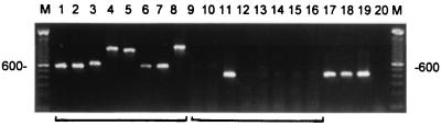
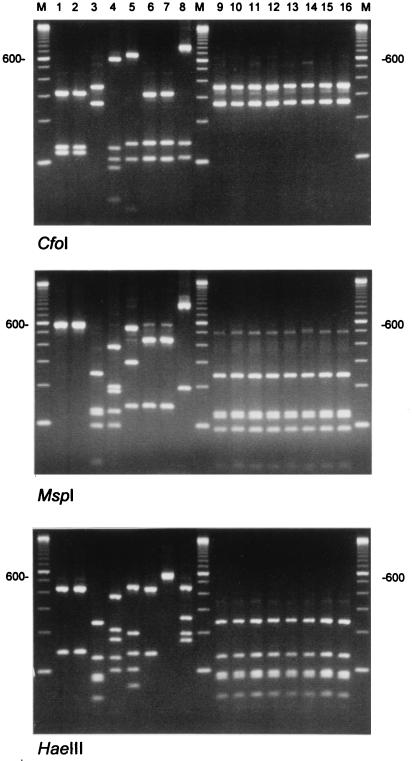
Madurella mycetomatis is the commonest cause of eumycetoma in Sudan and other countries in tropical Africa. Currently, the early diagnosis of mycetoma is difficult. In attempting to improve the identification of M. mycetomatis and, consequently, the diagnosis of mycetoma, we have developed specific oligonucleotide primers based on the sequence of the internal transcribed spacer (ITS) regions spacing the genes encoding the fungal ribosomal RNAs. The ITS regions were amplified with universal primers and sequenced, and then two sets of species-specific primers were designed which specifically amplify parts of the ITS and the 5.8S ribosomal DNA gene. The new primers were tested for specificity with DNA isolated from human mycetoma lesions and DNA extracted from cultures of M. mycetomatis reference strains and related fungi as well as human DNA. To study the genetic variability of the ITS regions of M. mycetomatis, ITS amplicons were obtained from 25 different clinical isolates and subjected to restriction fragment length polymorphism (RFLP) analysis with CfoI, HaeIII, MspI, Sau3AI, RsaI, and SpeI restriction enzymes. RFLP analysis of the ITS region did not reveal even a single difference, indicating the homogeneity of the isolates analyzed during the current study.


Figures

FIG. 1
FIG. 1
PCR amplification of the ITS for M. mycetomatis and related species. Lanes 1 to 8 show the amplicons obtained by using the universal primers ITS4 and ITS5. DNAs from the following species were amplified: M. grisea CBS331-50 (lane 1) and CBS 332-50 (lane 2), M. mycetomatis CBS 247.48 (lane 3) and CBS 868.95 (lane 4), P. mackinnonii CBS 674.75 (lane 5), P. romeri CBS 252.60 (lane 6), P. unguis-homini CBS 378.92 (lane 7), and C. larense CBS 640.73 (lane 8). In lanes 9 to 16 PCR results obtained with the primer combination 26.1A and 28.3A are displayed; the strains are ordered similarly from left to right. Note that only M. mycetomatis CBS 247.48 yielded a positive signal. Lanes 17 to 19 show the representative PCR products obtained for all of the clinical M. mycetomatis isolates. Lane 20 contains the negative PCR control. On the left and right the molecular size of the intensely fluorescing 600-bp-long fragment in the 100-bp ladder is indicated.
FIG. 2
FIG. 2
RFLP analysis of the ITS amplicons obtained for M. mycetomatis and related species. Lanes 1 to 8 show the results obtained for M. grisea CBS331-50 (lane 1) and CBS 332-50 (lane 2), M. mycetomatis CBS 247.48 (lane 3) and CBS 868.95 (lane 4), P. mackinnonii CBS 674.75 (lane 5), P. romeri CBS 252.60 (lane 6), P. unguis-homini CBS 378.92 (lane 7), and C. larense CBS 640.73 (lane 8). The three panels were generated with the restriction enzymes indicated. Lanes 9 to 16 show the results obtained for eight of the clinical M. mycetomatis isolates, indicating the high degree of ITS homogeneity. Lanes M contain the 100-bp size marker ladder; the position of the 600-bp-long fragment is indicated.

References

    1. Ahmed A O A, Abugroun E A M, Fahal A H, Zijlstra E E, van Belkum A, Verbrugh H A. Unexpected high prevalence of secondary bacterial infection in patients with mycetoma. J Clin Microbiol. 1998;36:850–851. - PMC - PubMed
    1. Baleiras Couto M M, Vogels J T, Hofstra H, Huis in ’t Veld J H, Vossen J M. Random amplified polymorphic DNA and restriction enzyme analysis of PCR amplified rDNA in taxonomy: two identification techniques for food borne yeasts. J Appl Bacteriol. 1995;79:525–535. - PubMed
    1. Boom R, Sol C J A, Salimans M M M, Jansen C L, Wertheim-van Dillen P M E, van der Noordaa J. Rapid and simple method for purification of nucleic acids. J Clin Microbiol. 1990;28:495–503. - PMC - PubMed
    1. De Hoog G S, Buiting A, Tan C S, Stroebel A B, Ketterings C, de Boer E J, Naafs B, Brimicombe R, Nohlmans-Paulssen M K E, Fabius G T J, Klokke A H, Visser L G. Diagnostic problems with imported cases of mycetoma in The Netherlands. Mycoses. 1993;36:81–87. - PubMed
    1. El Hag I A, Fahal A H, Khalil E A G. Fine needle aspiration cytology of mycetoma. Acta Cytol. 1996;40:461–464. - PubMed

MeSH terms

Associated data

LinkOut - more resources

Cite

AltStyle によって変換されたページ (->オリジナル) /