This site needs JavaScript to work properly. Please enable it to take advantage of the complete set of features!
Skip to main page content
U.S. flag

An official website of the United States government

Dot gov

The .gov means it’s official.
Federal government websites often end in .gov or .mil. Before sharing sensitive information, make sure you’re on a federal government site.

Https

The site is secure.
The https:// ensures that you are connecting to the official website and that any information you provide is encrypted and transmitted securely.

NIH NLM Logo
Log in
Access keys NCBI Homepage MyNCBI Homepage Main Content Main Navigation
. 1999 May;37(5):1385-92.
doi: 10.1128/JCM.37.5.1385-1392.1999.

Detection by enzyme immunoassay of serum immunoglobulin G antibodies that recognize specific Cryptosporidium parvum antigens

Affiliations

Detection by enzyme immunoassay of serum immunoglobulin G antibodies that recognize specific Cryptosporidium parvum antigens

J W Priest et al. J Clin Microbiol. 1999 May.

Abstract

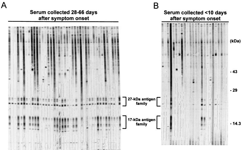
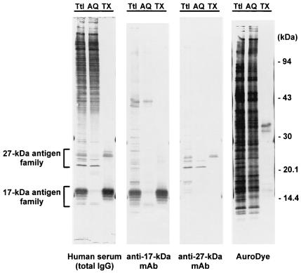
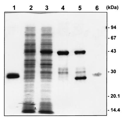
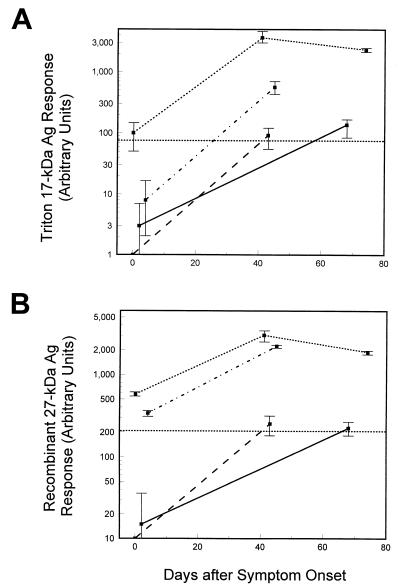
Human infection with Cryptosporidium parvum usually elicits characteristic immunoglobulin G (IgG), IgA, and IgM antibody responses against two sporozoite surface antigens with apparent molecular masses of approximately 27 and 17 kDa. We have determined that these two antigens are actually complex families of related antigens. We have developed two new enzyme-linked immunosorbent assays (ELISAs) for the detection and quantitation of serum IgG antibodies against both antigens. The assays utilize a recombinant form of the 27-kDa antigen and a partially purified native fraction isolated from sonicated whole oocysts that contains 17-kDa antigen. An immunoblot assay previously developed in our laboratory served as the reference, or "gold standard," seroassay for the assessment of the new ELISAs. Positive responses with the recombinant-27-kDa-antigen ELISA were correlated with the immunoblot results for the 27-kDa antigen, with a sensitivity and specificity of 90 and 92%, respectively. Similarly, positive responses with the partially purified native-17-kDa-antigen ELISA correlated with the immunoblot results for the 17-kDa antigen, with a sensitivity and specificity of 90 and 94%, respectively. For both ELISAs the median IgG antibody levels for serum sets collected during outbreaks of waterborne C. parvum infection were at least 2.5-fold higher than the levels determined for a nonoutbreak set. Using the immunoblot as the "gold standard," the new ELISAs were more specific and, in the case of the 27-kDa-antigen ELISA, more sensitive than the crude oocyst antigen ELISA currently in use. These assays will be useful in future epidemiologic studies.


Figures

FIG. 1
FIG. 1
Western blot of serum samples from symptomatic and asymptomatic Carrollton, Ga., residents. A crude antigen supernatant prepared from sonicated oocysts was resolved on an SDS–10 to 22.5% polyacrylamide gel under nonreducing conditions (600 ng/mm of gel width). Following electrotransfer to Immobilon P, 2-mm-wide strips of membrane were incubated overnight at 4°C with serum from individual patients with cryptosporidiosis at a dilution of 1:100 in 0.3% Tween 20-PBS. The blots were developed with a biotinylated anti-human IgG monoclonal antibody and alkaline phosphatase-labeled streptavidin as described in Materials and Methods. (A) Antigens recognized by serum collected from patients with cryptosporidiosis 28 to 66 days after symptom onset (late outbreak). (B) Antigens recognized by sera collected from patients less than 10 days after symptom onset (early outbreak). The locations of the 27- and 17-kDa-antigen families are indicated.
FIG. 2
FIG. 2
Extraction of 27- and 17-kDa antigens by Triton X-114. A crude antigen supernatant prepared from sonicated oocysts was fractionated by Triton X-114 phase partition extraction as described in Materials and Methods. Total unfractionated proteins (Ttl), proteins found in the detergent-depleted aqueous phase (AQ), and proteins extracted into the Triton X-114 detergent-rich phase (TX) were resolved by SDS-polyacrylamide gel electrophoresis as described in the legend to Fig. 1. The proteins were blotted onto Immobilon P and incubated overnight at 4°C with either a 1:100 dilution of human serum from a patient with cryptosporidiosis in 0.3% Tween 20-PBS (Human serum), a 1:1 dilution of tissue culture supernatant in 0.3% Tween 20-PBS containing a monoclonal antibody (C6C1) against the 17-kDa antigen (anti-17-kDa MAb), a 1:1 dilution of tissue culture supernatant in 0.3% Tween 20-PBS containing a monoclonal antibody (C6B6) against the 27-kDa antigen (anti-27-kDa MAb), or AuroDye-forte to stain all proteins (AuroDye). The blots were developed with either a biotinylated anti-human IgG monoclonal antibody and streptavidin-labeled alkaline phosphatase (human serum) or a horseradish peroxidase-labeled goat anti-mouse polyclonal antibody (anti-17-kDa MAb and anti-27-kDa MAb) as described in Materials and Methods. The positions of the molecular mass markers and the 27- and 17-kDa-antigen families are indicated.
FIG. 3
FIG. 3
Purification of recombinant 27-kDa protein. The protein sequence of the 27-kDa antigen was cloned into the pGEX expression system. Proteins from various stages of the expression and purification were resolved on an SDS–12% polyacrylamide gel and stained with Coomassie brilliant blue R-250. Lanes: (1) purified GST; (2) E. coli cells containing the plasmid construct before IPTG induction; (3) the same cells 4 h after IPTG induction; (4) purified fusion protein; (5) purified fusion protein after partial thrombin cleavage; (6) 2 μg of recombinant 27-kDa antigen after removal of uncleaved fusion protein and GST. Lane 6 was digitally enhanced to improve the visibility of the weakly staining recombinant 27-kDa protein band. The positions of the molecular mass markers are indicated.
FIG. 4
FIG. 4
ELISA responses for paired serum samples. Paired serum samples from patients with cryptosporidiosis were assayed for antigen (Ag)-specific IgG antibodies using the Triton antigen ELISA (A) or the recombinant-27-kDa-antigen ELISA (B) as described in Materials and Methods. The 76-arbitrary unit (A) and 206-unit (B) threshholds for positivity in the two ELISAs are indicated by dotted lines across the graphs. Samples with ELISA responses near or below this cutoff were assayed on three separate occasions, and all others were assayed twice. The mean values and 1 standard deviation are indicated. Each kind of line represents an individual patient.

References

    1. Arrowood M J. Ph.D. thesis. Tucson: University of Arizona; 1988.
    1. Arrowood M J, Mead J R, Mahrt J L, Sterling C R. Effects of immune colostrum and orally administered antisporozoite monoclonal antibodies on the outcome of Cryptosporidium parvum infections in neonatal mice. Infect Immun. 1989;57:2283–2288. - PMC - PubMed
    1. Arrowood M J, Sterling C R. Isolation of Cryptosporidium oocysts and sporozoites using discontinuous sucrose and isopycnic Percoll gradients. J Parasitol. 1987;73:314–319. - PubMed
    1. DuPont H L, Chappel C L, Sterling C R, Okhuysen P C, Rose J B, Jakubowski W. The infectivity of Cryptosporidium parvum in healthy volunteers. N Engl J Med. 1995;332:855–859. - PubMed
    1. Dworkin M S, Goldman D P, Wells T G, Kobayashi J M, Herwaldt B L. Cryptosporidiosis in Washington State: an outbreak associated with well water. J Infect Dis. 1996;174:1372–1376. - PubMed

MeSH terms

LinkOut - more resources

Cite

AltStyle によって変換されたページ (->オリジナル) /