This site needs JavaScript to work properly. Please enable it to take advantage of the complete set of features!
Skip to main page content
U.S. flag

An official website of the United States government

Dot gov

The .gov means it’s official.
Federal government websites often end in .gov or .mil. Before sharing sensitive information, make sure you’re on a federal government site.

Https

The site is secure.
The https:// ensures that you are connecting to the official website and that any information you provide is encrypted and transmitted securely.

NIH NLM Logo
Log in
Access keys NCBI Homepage MyNCBI Homepage Main Content Main Navigation
. 2025 Jan;262(1):207-218.
doi: 10.1007/s00709-024-01990-7. Epub 2024 Sep 23.

Chloroplasts with clefts and holes: a reassessment of the chloroplast shape using 3D FE-SEM cellular reconstruction of two species of Chlamydomonas

Affiliations

Chloroplasts with clefts and holes: a reassessment of the chloroplast shape using 3D FE-SEM cellular reconstruction of two species of Chlamydomonas

Naoki Sato et al. Protoplasma. 2025 Jan.

Abstract

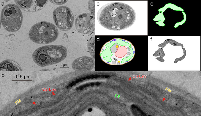
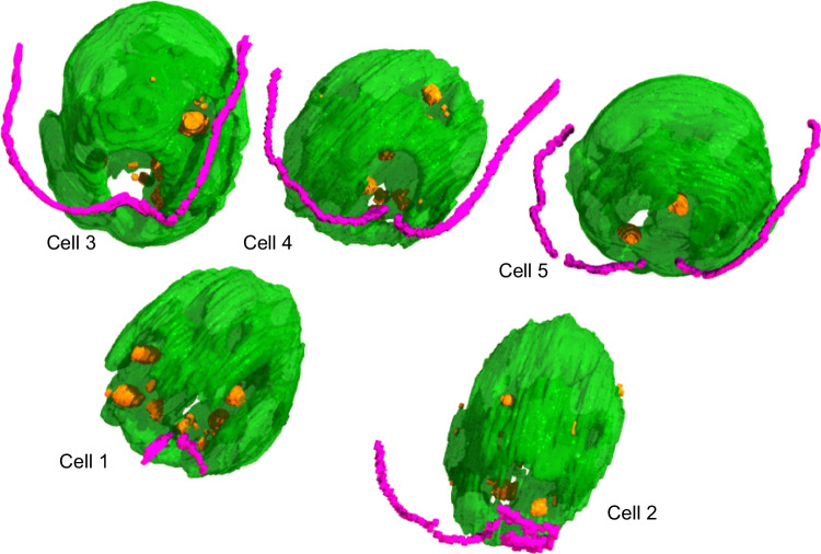
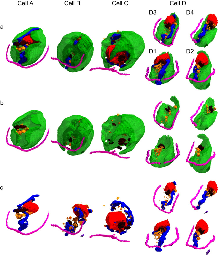
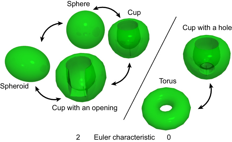
Chloroplasts are usually considered spheroid organelles, but this is not the only shape of chloroplasts. The chloroplast of Chlamydomonas has been typically described as cup-shaped. However, in old studies, it was also modeled as a complex shape with "perforations" or windows. Here, we reconstructed the cellular architecture of Chlamydomonas reinhardtii and C. applanata using an array tomography system installed on a field emission scanning electron microscope. C. reinhardtii chloroplasts resembled a baseball glove or a cup without a side, featuring numerous large and small holes that may facilitate the transport of metabolites and proteins produced in the Golgi apparatus fitted in the holes. In a lipid-accumulating, high-light condition, the chloroplast volume increased by filling the side cleft with an entire wall. Many accumulated large lipid droplets were accommodated within the chloroplast holes, which could have been considered as "chloroplast lipid droplets." Mitochondrial meshworks surrounded the chloroplast. C. applanata chloroplasts appeared like a folded starfish or a cup with many side clefts and a few holes. There was a single mitochondrion or two that branched in a complex form. Tight contacts of various organelles were also found in C. applanata. These reconstructions illustrate the complexity of chloroplast shape, which necessitates a revised understanding of the localization of lipid droplets and the evolution of chloroplasts: The prevailing image of the spheroid chloroplasts that reminds us of the similarity between chloroplasts and cyanobacteria is no longer tenable.

Keywords: Chlamydomonas; Array tomography; Chloroplast shape; Lipid droplet topology; Three-dimensional reconstruction.


Conflict of interest statement

Declarations. Competing interests: The authors declares no competing interests.

Figures

Fig. 1
Fig. 1
Sample image of chloroplast envelope and the flow of image processing. a A FE-SEM image of C. reinhardtii cells grown in the standard condition. b Enlargement of a part of the cell in the center of panel a. Cp, chloroplast; CpEnv (with a red arrow), chloroplast envelope membranes; PM, plasma membrane. c–f The flow of image processing. c Cropped image of the central cell in panel a. d The same cell with organelles delineated and colored ("color-painted organelles"). e Isolated chloroplast drawing. f Clipped chloroplast SEM image using the drawing in e as a mask. The clipped chloroplast SEM image was used for 3D reconstruction by the Fluorender software, whereas the color-painted chloroplast drawings (namely, without internal structures) were used for 3D reconstruction by the 3D Slicer software
Fig. 2
Fig. 2
Different views of the C. reinhardtii chloroplasts of three cells (Cell 1, Cell 2, and Cell 3) grown in the standard condition. The 3D views were reconstructed from the multi-TIF files for the clipped chloroplast SEM images and the color-painted drawings of the other organelles with the Fluorender software. The structures other than chloroplast are not shown in all panels. All the images show perspective views with similar scaling except in panel (l), which is an enlarged image to show small holes. Top (a, e, i), anterior view, namely, the view from the flagella side. The black arrow indicates the large opening of the chloroplast. Second row (b, f, j), side view from the large opening showing the holes in the opposite chloroplast lobe. Third row (c, g, k), view with clipping to show the pyrenoids (red arrows). Bottom (d, h, l), view from the chloroplast lobe to show small holes. Green, chloroplast; purple, flagella; red, nucleus; blue, mitochondrion; orange, lipid droplet; dark gray, Golgi apparatus. Cell wall, plasma membrane, and vacuoles are not included in the figures to show the internal structure of the cell
Fig. 3
Fig. 3
3D structures of the C. reinhardtii cells grown in the lipid-accumulating condition. The 3D surface models created by the 3D Slicer software were assembled using the Shade 3D software. Not all parts of flagella were modeled. For color codes, see the legend for Fig. 2
Fig. 4
Fig. 4
Comparison of the C. reinhardtii cells grown in the standard (left) and lipid-accumulating (right) conditions. Top (a, c), whole cell view without cell wall, plasma membrane and vacuoles. Bottom (b, d), view without chloroplast. For color codes, see the legend for Fig. 2
Fig. 5
Fig. 5
Comparison of the volumes of various cellular parts of C. reinhardtii grown in the standard (blue) and lipid-accumulating (orange) conditions. The volumetry was based on the STL model constructed by the 3D Slicer software. Each error bar indicates the standard deviation (n = 3 for the standard condition, n = 5 for the lipid-accumulating condition)
Fig. 6
Fig. 6
3D structures of C. applanata cells. Four cells are arranged according to putative progress of cell cycle. a Whole cell view without cell wall, plasma membrane, and vacuoles. b View of chloroplast and flagella with lipid droplets and Golgi apparatuses. c View as a but without chloroplast. Cell D consisted of four daughter cells, D1, D2, D3, and D4. Dark purple, eye spot. For other color codes, see the legend for Fig. 2
Fig. 7
Fig. 7
Homeomorphism of closed surfaces. Sphere, spheroid, cup, and cup with an opening are homeomorphic, having the Euler characteristic of 2. Torus and cup with a hole are homeomorphic, having the Euler characteristic of 0. The presence of a hole implies the importance of negative curvature and thus introduces a fundamental difference in both mathematics and biology

References

    1. Deason TR, Bold HC (1960) Phycological Studies I. The University of Texas Publication, Exploratory studies on Texas soil algae
    1. Engel BD, Schaffer M, Cuellar LK, Villa E, Plitzko JM, Baumeister W (2015) Native architecture of the Chlamydomonas chloroplast revealed by in situ cryo-electron tomography eLife 4:e04889 10.7554/eLife.04889 - PMC - PubMed
    1. Ettl H, Schlösser UG (1992) Towards a revision of the systematics of the genus Chlamydomonas (Chlorophyta). I Chlamydomonas Applanata Pringsheim Bot Acta 105:323–330
    1. Feng L, Guo W, Guo J, Zhang X, Zou X, Rao M, Ye J, Kuang C, Chen G, Chen C, Qin S, Yang W, Cheng J (2023) FIB-SEM analysis on three-dimensional structures of growing organelles in wild Chlorellapyrenoidosa cells. Protoplasma 260:885–897. 10.1007/s00709-022-01821-7 - PubMed
    1. Freeman Rosenzweig ES, Xu B, Kuhn Cuellar L, Martinez-Sanchez A, Schaffer M, Strauss M, Cartwright HN, Ronceray P, Plitzko JM, Förster F, Wingreen NS, Engel BD, Mackinder LCM, Jonikas MC (2017) The eukaryotic CO2-concentrating organelle is liquid-like and exhibits dynamic reorganization. Cell 171:148–162. 10.1016/j.cell.201708008 - PMC - PubMed

MeSH terms

LinkOut - more resources

Cite

AltStyle によって変換されたページ (->オリジナル) /