This site needs JavaScript to work properly. Please enable it to take advantage of the complete set of features!
Skip to main page content
U.S. flag

An official website of the United States government

Dot gov

The .gov means it’s official.
Federal government websites often end in .gov or .mil. Before sharing sensitive information, make sure you’re on a federal government site.

Https

The site is secure.
The https:// ensures that you are connecting to the official website and that any information you provide is encrypted and transmitted securely.

NIH NLM Logo
Log in
Access keys NCBI Homepage MyNCBI Homepage Main Content Main Navigation
. 2022 May 25;144(20):9023-9032.
doi: 10.1021/jacs.2c01401. Epub 2022 May 13.

Widespread Sterol Methyltransferase Participates in the Biosynthesis of Both C4α- and C4β-Methyl Sterols

Affiliations

Widespread Sterol Methyltransferase Participates in the Biosynthesis of Both C4α- and C4β-Methyl Sterols

Wenxu Zhou et al. J Am Chem Soc. .

Abstract

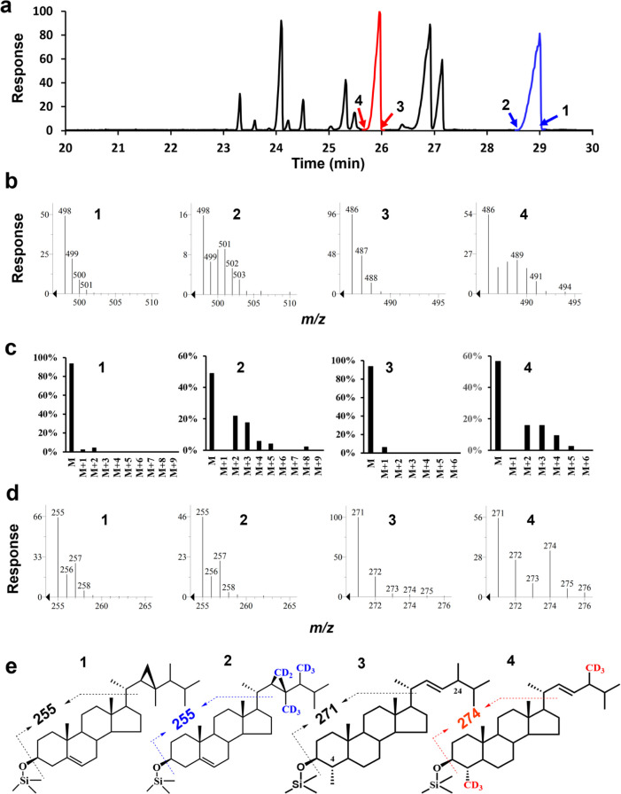
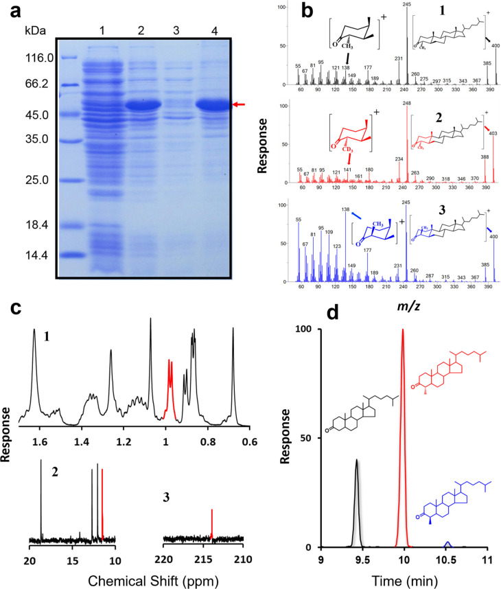
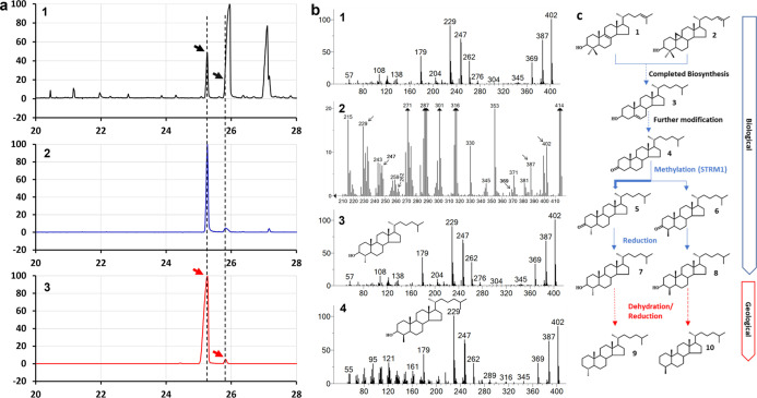
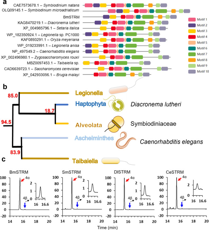
The 4-methyl steranes serve as molecular fossils and are used for studying both eukaryotic evolution and geological history. The occurrence of 4α-methyl steranes in sediments has long been considered evidence of products of partial demethylation mediated by sterol methyl oxidases (SMOs), while 4β-methyl steranes are attributed entirely to diagenetic generation from 4α-methyl steroids since possible biological sources of their precursor 4β-methyl sterols are unknown. Here, we report a previously unknown C4-methyl sterol biosynthetic pathway involving a sterol methyltransferase rather than the SMOs. We show that both C4α- and C4β-methyl sterols are end products of the sterol biosynthetic pathway in an endosymbiont of reef corals, Breviolum minutum, while this mechanism exists not only in dinoflagellates but also in eukaryotes from alveolates, haptophytes, and aschelminthes. Our discovery provides a previously untapped route for the generation of C4-methyl steranes and overturns the paradigm that all 4β-methyl steranes are diagenetically generated from the 4α isomers. This may facilitate the interpretation of molecular fossils and understanding of the evolution of eukaryotic life in general.


Conflict of interest statement

The authors declare no competing financial interest.

Figures

Figure 1
Figure 1
Sterol profiles of B. minutum incubated with [2H3-methyl]-methionine. (a) GC trace of sterol trimethylsilyl ethers. The red peak is 4,24-dimethyl-5α-cholest-22-en-3β-ol, while the blue peak is gorgosterol. The arrows indicate mass spectra of the nondeuterated (1,3) and deuterated (2,4) sterols. (b) Partial mass spectra of substances at the leading and trailing edges of the peaks, indicating differences in their degrees of deuterium incorporation of unlabeled gorgosterol (1), labeled gorgosterol (2), unlabeled 4,24-dimethyl-5α-cholest-22E-en-3β-ol (3), and labeled 4,24-dimethyl-5α-cholest-22E-en-3β-ol (4). (c) Isotopic pattern deconvolution of the mass spectra of unlabeled gorgosterol (1), labeled gorgosterol (2), unlabeled 4α,24-dimethyl-5α-cholest-22E-en-3β-ol (3), and labeled 4α,24-dimethyl-5α-cholest-22E-en-3β-ol (4). (d) Partial mass spectra of the nuclei of unlabeled gorgosterol (1), labeled gorgosterol (2), unlabeled 4α,24-dimethylcholest-22E-en-3β-ol (3), and labeled 4α,24-dimethylcholest-22E-en-3β-ol (4). (e) Structures of unlabeled gorgosterol (1), labeled gorgosterol (2), unlabeled 4,24-dimethyl-5α-cholest-22E-en-3β-ol (3), and labeled 4α,24-dimethylcholest-22E-en-3β-ol (4).
Figure 2
Figure 2
In vitro assays of BmSTRM enzymatic activity. (a) Visualization, by gel imaging, of BmSTRM protein expressed in E. coli and separated in a precast 12% Bis-Tris gel. Lane 1, control with no induction by IPTG; Lane 2, proteins of broken cells induced by IPTG; Lanes 3–4, soluble (3) and insoluble fractions (4) of proteins of BmSTRM-expressing E. coli. (b) Comparison of mass spectra of 4α-methylcholestanone products obtained from reactions with BmSTRM, cholestanone, and AdoMet (1) or [2H3-methyl] AdoMet (2). Comparison of the mass spectra of 4α- and 4β-methylcholestanone (3). (c) PNMR spectroscopy and 13C NMR analysis of purified BmSTRM products. (1) PNMR spectroscopic analysis of the 4α-methyl group (with a doublet signal centered at 0.98 ppm), (2) 13C NMR analysis of the diagnostic signals of the 4α-methyl group at 11.5 ppm, and (3) 13C NMR analysis of the attachment point of the keto group at 214 ppm. (d) GC trace of products of the in vitro BmSTRM assay with a reaction mixture of cholestanone and 750 μM AdoMet.
Figure 3
Figure 3
GC–MS chromatographic and spectroscopic analysis of 4-methycholestanol obtained from B. minutum. (a) Chromatograms of fraction 41 obtained from HPLC separation of sterols (see Results for details) of B. minutum in a full-scan mode, with arrows indicating peaks of 4α-methylcholestanol (lophanol) and 4β-methylcholestanol (1); fraction 41 in SIM mode using m/z 402 (2); and generated reference standards (4-methylated products of the BmSTRM-catalyzed reaction in the in vitro feeding assay) in the SIM mode using m/z 402 (3). Note: the dotted lines indicate identical retention times in chromatograms. (b) Mass spectra of the following substances in fraction 41: 4α-methylcholestanol (1); the fraction’s component with a GC–MS retention time of 25.27 min and diagnostic fragments of 4β-methylcholestanol indicated by arrows (2); and authentic 4α-methylcholestanol and 4β-methylcholestanol derived from C4-methylated products of the BmSTRM-catalyzed reaction in the in vitro feeding assay with cholestanone as substrate (3 and 4, respectively). (c) Deduced route of 4-methylsterane generation. Proportions of the 4-methylated isomers indicate that sterols in fossil records in immature sediments have biogenic sources and conserved configurations rather than originating from geological processes. (1) lanosterol; (2) cycloartenol; (3) cholesterol; (4) cholestanone; (5) 4α-methylcholestanone; (6) 4β-methylcholestanone; (7) 4α-methylcholestanol; (8) 4β-methylcholestanol; (9) 4α-methylsterane; and (10) 4β-methylsterane.
Figure 4
Figure 4
Phylogenetic analysis and enzymatic activity of STRMs from S. microadriaticum, D. lutheri, and C. elegans. (a) Illustration of conserved motifs among the representative species. Different colors of the boxes refer to different motifs from 1 to 10. (b) Origin and diversification of the STRM gene family. Average divergence time are indicated for nodes of interest (million years, Ma). See Figure S6 for details. (c) Comparison of the enzymatic activity of BmSTRM and the STRMs of S. microadriaticum (SmSTRM), D. lutheri (DlSTRM), and C. elegans (CeSTRM). Chromatograms of products generated by BmSTRM (1), SmSTRM (2), DlSTRM (3), and CeSTRM (4) in a full-scan mode, with arrows indicating peaks of 4α-methylcholestanol (lophenol; the red arrows) and 4β-methylcholestanol (the blue arrows). Note: the dotted lines indicate enlarged chromatograms of the 4β isomer. See Figure S7 for the mass spectra of 4α- and 4β-methylcholestanol produced by SmSTRM, DlSTRM, and CeSTRM.

References

    1. Pollier J.; et al. A widespread alternative squalene epoxidase participates in eukaryote steroid biosynthesis. Nat. Microbiol. 2018, 4, 226.10.1038/s41564-018-0305-5. - DOI - PubMed
    1. Kodner R. B.; Summons R. E.; Pearson A.; King N.; Knoll A. H. Sterols in a unicellular relative of the metazoans. Proc. Natl. Acad. Sci. U.S.A. 2008, 105, 9897–9902. 10.1073/pnas.0803975105. - DOI - PMC - PubMed
    1. Goad L. J. The biosynthesis of plant sterols. Lipids Lipid Polym. Higher Plants, [Pap. Symp.] 1977, 146–168. 10.1007/978-3-642-66632-2_8. - DOI
    1. Knapp F. F.; Trowbridge S. T.; Schroepfer G. J. Concerning the role of 4.beta.-methyl sterols in cholesterol biosynthesis. J. Am. Chem. Soc. 1975, 97, 3522–3524. 10.1021/ja00845a044. - DOI - PubMed
    1. Robinson N.; Eglinton G.; Brassell S. C.; Cranwell P. A. Dinoflagellate origin for sedimentary 4α-methylsteroids and 5α(H)-stanols. Nature 1984, 308, 439–442. 10.1038/308439a0. - DOI

Publication types

Cite

AltStyle によって変換されたページ (->オリジナル) /