This site needs JavaScript to work properly. Please enable it to take advantage of the complete set of features!
Skip to main page content
U.S. flag

An official website of the United States government

Dot gov

The .gov means it’s official.
Federal government websites often end in .gov or .mil. Before sharing sensitive information, make sure you’re on a federal government site.

Https

The site is secure.
The https:// ensures that you are connecting to the official website and that any information you provide is encrypted and transmitted securely.

NIH NLM Logo
Log in
Access keys NCBI Homepage MyNCBI Homepage Main Content Main Navigation
. 2020 Dec 22;117(51):32722-32730.
doi: 10.1073/pnas.2005460117. Epub 2020 Dec 8.

Guanine, a high-capacity and rapid-turnover nitrogen reserve in microalgal cells

Affiliations

Guanine, a high-capacity and rapid-turnover nitrogen reserve in microalgal cells

Peter Mojzeš et al. Proc Natl Acad Sci U S A. .

Abstract

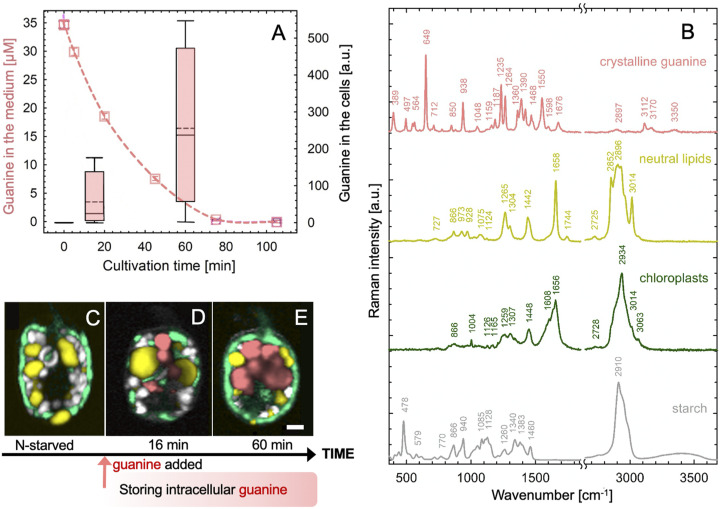
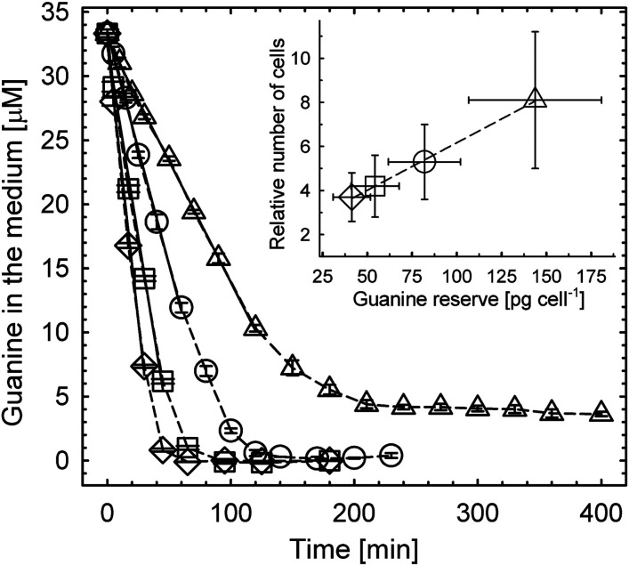
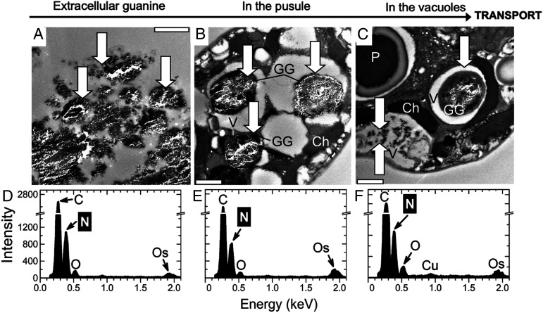
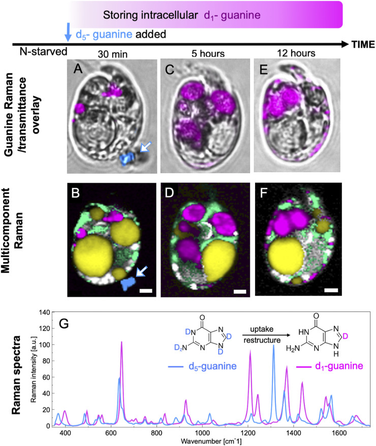
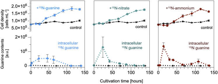
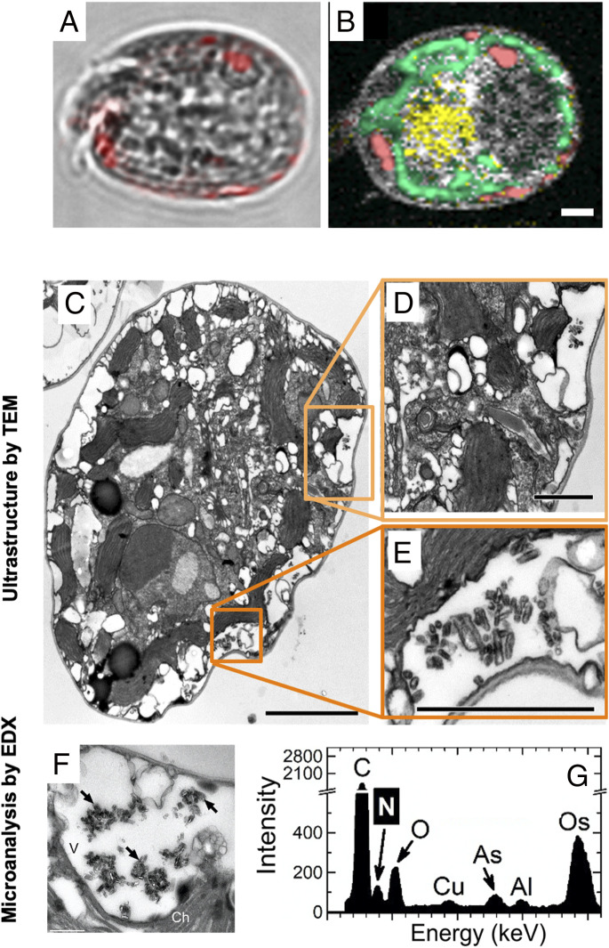
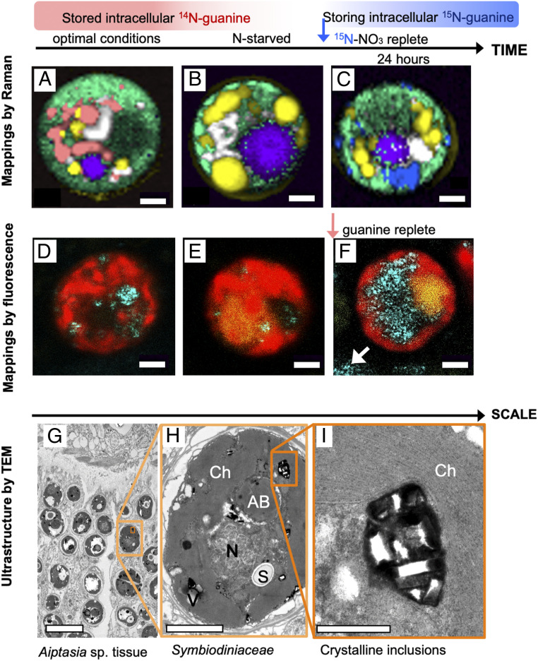
Nitrogen (N) is an essential macronutrient for microalgae, influencing their productivity, composition, and growth dynamics. Despite the dramatic consequences of N starvation, many free-living and endosymbiotic microalgae thrive in N-poor and N-fluctuating environments, giving rise to questions about the existence and nature of their long-term N reserves. Our understanding of these processes requires a unequivocal identification of the N reserves in microalgal cells as well as their turnover kinetics and subcellular localization. Herein, we identified crystalline guanine as the enigmatic large-capacity and rapid-turnover N reserve of microalgae. The identification was unambiguously supported by confocal Raman, fluorescence, and analytical transmission electron microscopies as well as stable isotope labeling. We discovered that the storing capacity for crystalline guanine by the marine dinoflagellate Amphidiniumcarterae was sufficient to support N requirements for several new generations. We determined that N reserves were rapidly accumulated from guanine available in the environment as well as biosynthesized from various N-containing nutrients. Storage of exogenic N in the form of crystalline guanine was found broadly distributed across taxonomically distant groups of microalgae from diverse habitats, from freshwater and marine free-living forms to endosymbiotic microalgae of reef-building corals (Acropora millepora, Euphyllia paraancora). We propose that crystalline guanine is the elusive N depot that mitigates the negative consequences of episodic N shortage. Guanine (C5H5N5O) may act similarly to cyanophycin (C10H19N5O5) granules in cyanobacteria. Considering the phytoplankton nitrogen pool size and dynamics, guanine is proposed to be an important storage form participating in the global N cycle.

Keywords: coral; guanine; nitrogen cycle; nutrient storage; phytoplankton.


Conflict of interest statement

The authors declare no competing interest.

Figures

Fig. 1.
Fig. 1.
Rapid uptake of guanine by N-starved A. carterae led to the accumulation of intracellular guanine inclusions. Cells were suspended in a saturated solution of guanine (∼35 μM at 20 °C) in N-deficient f/2 medium at a density of 1.7 ± 0.3 ×ばつ 105 cells·mL−1. The dashed line in A shows the declining concentration of guanine in the medium as measured by ultraviolet absorption (SI Appendix, section II.1.B). The simultaneous accumulation of guanine in the cells was assessed using Raman microscopy (SI Appendix, section II.1.C) and is shown in the boxplot. Raman spectra in B were used to generate the Raman maps in CE that represent: (C) a typical cell after 2 wk without a N source and (D and E) cells during progressive guanine accumulation. Color legend: guanine (pink), lipids (yellow), chloroplast (green), and starch (white/gray). (Scale bar, 2 μm.) Raman maps showing separate cellular constituents in CE are constructed as described in SI Appendix, section I.1 and presented in SI Appendix, Fig. S1.
Fig. 2.
Fig. 2.
Uptake of dissolved guanine from the medium by N-starved A. carterae and the number of generations supported by accumulated reserves (inset). The rate of guanine disappearance from the medium decreased with reduced cell density (±SD): from (122 ± 31) (◇) to (93 ± 23) (しろいしかく), to (61 ± 15) (◯), and to (31 ± 8) (しろさんかく) ×ばつ 103 cells·mL−1. The most dilute culture (しろさんかく) reduced guanine concentration in the medium from 35 to ∼4 μM, revealing the maximum storage capacity of cells of 143 ± 37 (SD) pg of crystalline guanine cell−1. Inset shows the correlation between the number of cells grown on guanine reserve and the reserve size. Details of calculations for this figure are described in SI Appendix, section I.2 and Fig. S2.
Fig. 3.
Fig. 3.
TEM of semithin sections (AC) and energy-dispersive X-ray spectroscopy (EDX) (D–F) analysis of A. carterae 6 h after refeeding N-starved cultures with guanine. Typical EDX point spectra of guanine crystals (DF) indicating a high N content were obtained in the scanning TEM (STEM) mode from semithin cell sections. Arrows point to guanine particles outside (A) and inside cells (B and C). (Scale bars, 1 μm.) Ch, chloroplast; GG, globules with microcrystalline guanine; P, pyrenoid; V, vacuole.
Fig. 4.
Fig. 4.
Uptake of guanine includes exchange of deuterium for hydrogen atoms. Bright-field images overlaid by guanine (A, C, and E) and multicomponent Raman maps (B, D, and F) of N-starved A. carterae after the addition of solid crystalline fully deuterated d5-guanine to N-depleted medium. Images collected 30 min (A and B), 5 h (C and D), and 12 h (E and F) after d5-guanine addition. Data for d5-guanine and partially deuterated d1-guanine are presented in blue and magenta, respectively, in both the Raman spectrum and the images. G shows their respective Raman spectra. Other colors: yellow, neutral lipids; green, chloroplasts; white/gray, starch. (Scale bars [AF], 2 μm.) More spectra of isotopically labeled guanine are shown in SI Appendix, Fig. S7. Raman maps showing separate components from data represented in B, D, and F are provided in SI Appendix, Fig. S9.
Fig. 5.
Fig. 5.
N in guanine inclusions originated directly from the supplied guanine, nitrate, and ammonium. A. carterae cell density stagnated in controls without N feeding (black lines) and divided after addition of 15N-labeled guanine, nitrate, and ammonium (all at 0.882-mM N). Intracellular crystalline guanine per cell is shown in the bottom graphs representing Raman measurements (n = 5–12 cells). The corresponding graph representing uptake of urea is shown in SI Appendix, Fig. S10.
Fig. 6.
Fig. 6.
Localization of guanine inclusions in A. carterae fed by nitrate by TEM. Bright-field images overlaid by guanine (pink) (A) and multicomponent Raman maps (B) of A. carterae cells 24 h after refeeding N-starved cells with nitrate. False color coding is the same as that in Fig. 1. (Scale bar, 2 μm.) TEM of ultrathin (CE) and semithin (F) sections as well as EDX (G); (F and G) analysis of A. carterae cells and surrounding cells 26 h after refeeding N-starved cultures with nitrate. Typical EDX point spectra of guanine crystals (G) indicating high N content were obtained in the STEM mode from semithin cell sections. Arrows point to guanine crystals. (Scale bars, 2.5 [C], 1 [D and E], and 0.5 μm [F]. Ch, chloroplast; V, vacuole.)
Fig. 7.
Fig. 7.
Guanine in the endosymbiotic Symbiodiniaceae cells in corals. Raman maps of cells from tissue of the coral E. paraancora (AC). The spectra used to construct these maps are shown in SI Appendix, Fig. S13. Symbionts in corals maintained under optimal nutrient conditions contained guanine crystals (A). Cells from corals that were kept in N-depleted seawater for 4 mo contained very few guanine crystals (B). Twenty-four hours after feeding the starved coral 0.3-mM 15N-NaNO3, cells showed large 15N-guanine depots (C). The false color coding is the same as that in Figs. 1 and 4 with magenta added to represent accumulation bodies. (Scale bar, 2 μm.) Symbiodiniaceae cells isolated from the Great Barrier Reef (GBR) coral, A. millepora (DF). Cell from freshly collected coral (D), N-depleted coral (E), and from one day after feeding with medium containing traces of undissolved guanine grains (F). Cyan, 488-nm laser reflection of guanine grains outside (white arrow) and inside the cells; red, chlorophyll autofluorescence at 670–700 nm; yellow, fluorescence in accumulation bodies at 500–560 nm; white arrow, remains of undissolved crystalline guanine in medium. (Scale bar, 2 μm.) Ultrastructure of Symbiodiniaceae cells in Aiptasia sp. shown at increasing magnification (GI). AB, accumulation body; Ch, chloroplast; N, nucleus; S, floridean starch; V, vacuole. (Scale bars, 10 [G], 2 [H], and 0.5 μm [I].)

References

    1. Antia N. J., Harrison P. J., Oliveira L., The role of dissolved organic nitrogen in phytoplankton nutrition, cell biology and ecology. Phycologia 30, 1–89 (1991).
    1. Falkowski P. G., Evolution of the nitrogen cycle and its influence on the biological sequestration of CO2 in the ocean. Nature 387, 272–275 (1997).
    1. Falkowski P. G., Barber R. T., Smetacek V., Biogeochemical controls and feedbacks on ocean primary production. Science 281, 200–207 (1998). - PubMed
    1. Anderson D. M., Glibert P. M., Burkholder J. M., Harmful algal blooms and eutrophication: Nutrient sources, composition, and consequences. Estuaries 25, 704–726 (2002).
    1. Rädecker N., Pogoreutz C., Voolstra C. R., Wiedenmann J., Wild C., Nitrogen cycling in corals: The key to understanding holobiont functioning? Trends Microbiol. 23, 490–497 (2015). - PubMed

Publication types

LinkOut - more resources

Cite

AltStyle によって変換されたページ (->オリジナル) /