This site needs JavaScript to work properly. Please enable it to take advantage of the complete set of features!
Skip to main page content
U.S. flag

An official website of the United States government

Dot gov

The .gov means it’s official.
Federal government websites often end in .gov or .mil. Before sharing sensitive information, make sure you’re on a federal government site.

Https

The site is secure.
The https:// ensures that you are connecting to the official website and that any information you provide is encrypted and transmitted securely.

NIH NLM Logo
Log in
Access keys NCBI Homepage MyNCBI Homepage Main Content Main Navigation
. 2019 Dec 31;9(1):20416.
doi: 10.1038/s41598-019-56882-1.

Culture-independent and culture-dependent analyses of the bacterial community in the phycosphere of cyanobloom-forming Microcystis aeruginosa

Affiliations

Culture-independent and culture-dependent analyses of the bacterial community in the phycosphere of cyanobloom-forming Microcystis aeruginosa

Minkyung Kim et al. Sci Rep. .

Abstract

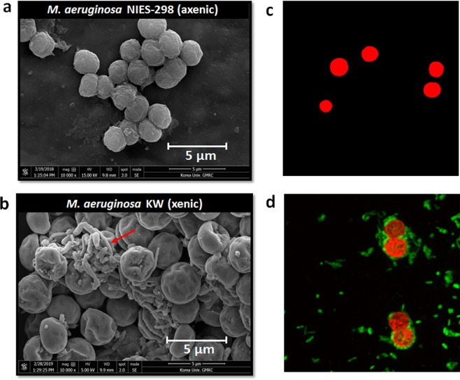
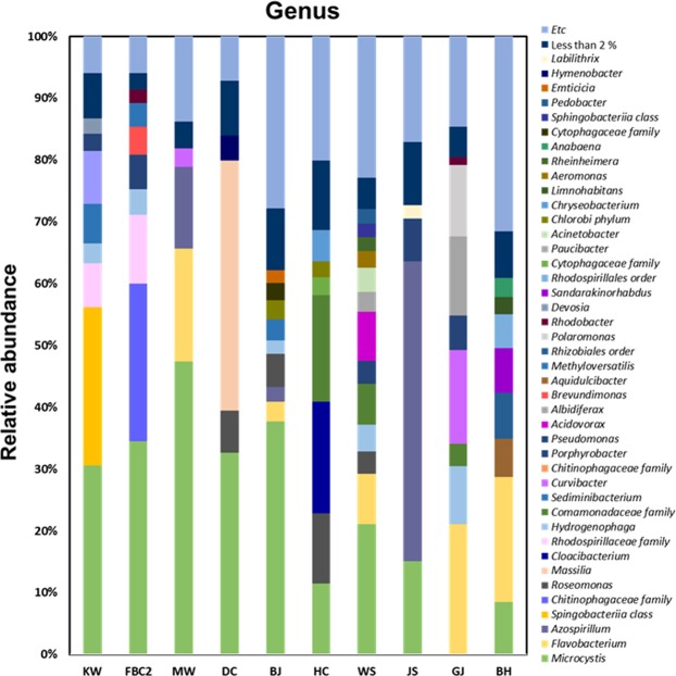
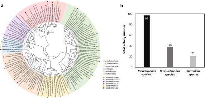
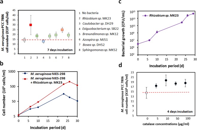
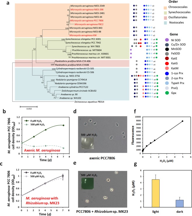
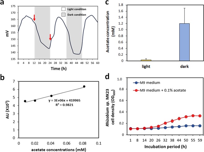
Confocal and scanning electron microscopic observations have previously shown the strong bacterial association of Microcystis aeruginosa cells on their surfaces. DNA-based analyses of the associated bacterial communities were carried out using two M. aeruginosa strains grown in the laboratory and eight newly collected cyanobacterial bloom samples. M. aeruginosa was the most predominant species (66-100%) within the phylum Cyanobacteria. Rhizobium, Hydrogenophaga and Brevundimonas species were commonly found, and Flavobacterium species were present in all the cyanobacterial bloom samples. In total, 396 colonies from various samples were screened, revealing that most culturable bacteria belonged to the class Alphaproteobacteria (19%) including Rhizobium, Brevundimonas, and Porphyrobacter species. The genetic variation among the M. aeruginosa strains and different habitat conditions may have led to the presence of distinct bacterial populations among the tested samples. Among all the tested seven culturable isolates, Rhizobium sp. MK23 showed the best growth-promotion effect on the axenic M. aeruginosa strains. H2O2 was observed to be produced during the growth of M. aeruginosa PCC7806 under light conditions, this strain was more resistant to H2O2 when associated with Rhizobium sp. MK23. Our data suggested that Rhizobium species along with other associated bacteria might help the growth of M. aeruginosa by decomposing H2O2 under the aerobic growing conditions.


Conflict of interest statement

The authors declare no competing interests.

Figures

Figure 1
Figure 1
Microcystis-bacteria co-existence. Scanning electron microscopy (SEM) image of the (a) axenic M. aeruginosa NIES-298 culture, and (b) xenic M. aeruginosa KW. SYTOTM 9 staining analysis of the (c) axenic M. aeruginosa NIES-298 and (d) xenic M. aeruginosa KW. The red and green represents M. aeruginosa and bacteria, respectively. The bacteria associated with the M. aeruginosa surface.
Figure 2
Figure 2
Bacterial community analysis of all the following M. aeruginosa samples: M. aeruginosa KW (KW), M. aeruginosa FBC000002 (FBC2), Murwang (MW), Daecheong (DC), Baekje (BJ), Hapcheon (HC), Wangsong (WS), Juksan (JS), Gangjeong (GJ), and Bohyun (BH). The genera with <1% abundance were included as well. The laboratory-cultured samples consisted of simple bacterial communities, whereas the environmental samples had various bacterial populations.
Figure 3
Figure 3
Culture-dependent analysis of the 16S rRNA gene sequences. (a) Neighbor‐joining phylogenetic tree of the isolated bacteria. Each color represents a phylum level of the bacterial community. The associated bacteria were isolated from KW, FBC2, MW, DC, BJ and HC samples. The number in parentheses indicates the number of isolated associated bacteria. Most bacteria belonged to the class Alpha-proteobacteria. GenBank accession numbers are provided in parentheses. (b) Pseudomonas species were the most frequently isolated species from the culturable bacteria.
Figure 4
Figure 4
(a) Co-culture experiments of M. aeruginosa PCC7806 with seven genera, Rhizobium sp. MK23 apparently promoted the growth of M. aeruginosa PCC7806 for 7 days. **p < 0.05. (b) M. aeruginosa NIES-298 grew faster with Rhizobium sp. MK23. The M. aeruginosa NIES-298 culture and co-culture started degrading on 23 and 26, repectively. (c) Rhizobium sp. MK23 in co-culture grew at a density of 1014 cells/ml in 30 days. (d) Addition of catalase promoted the growth of M. aeruginosa PCC7806 for 4 days.
Figure 5
Figure 5
M. aeruginosa cells are sensitive to oxidative stress. (a) The listed cyanobacterial antioxidant genes are SOD, superoxide dismutase; Kat, catalase; Prx, peroxidase; and Gpx, glutathione peroxidase. M. aeruginosa does not have a gene for producing a catalase. (b) Axenic of M. aeruginosa PCC7806 and M. aeruginosa PCC7806 with Rhizobium sp. MK23 were treated with 500 μM concentration of H2O2. After 7 days of treatment, the growth of M. aeruginosa PCC7806 was retarded under H2O2. (c) The growth of axenic M. aeruginosa PCC7806 with Rhizobium sp. MK23 did not change significantly. (d) The axenic PCC7806 treated with H2O2 was transparent in color and had an unusual cell shape. (e) PCC7806 co-cultured with Rhizobium sp. MK23 were still green and had a normal, round shape. (f) The standard curve for H202 concentrations in H2O2 assay. (g) In the H2O2 assay, the concentrations of H2O2 were much higher in the light condition than in dark.
Figure 6
Figure 6
(a) Dissolved oxygen (DO) changes during the growth of M. aeruginosa PCC7806 under the light and dark conditions for 60 h. In M. aeruginosa PCC7806 cultures, DO increased in the light condition and decreased in the dark condition. Cell supernatant was extracted at 12 h and 24 h (red arrows). (b) The standard curve for the acetate concentrations in HPLC analysis. (c) The HPLC analysis results showed that the concentration of acetate was much higher in the dark condition than in the light. (d) Growth curve of Rhizobium sp. MK23 under 0.1% acetate.

References

    1. Rabalais NN, et al. Dynamics and distribution of natural and human-caused hypoxia. Biogeosciences. 2010;7:585–619. doi: 10.5194/bg-7-585-2010. - DOI
    1. Paerl HW, Otten TG. Harmful cyanobacterial blooms: causes, consequences, and controls. Microb. Ecol. 2013;65:995–1010. doi: 10.1007/s00248-012-0159-y. - DOI - PubMed
    1. Schindler DW. Evolution of phosphorus limitation in lakes. Science. 1977;195:260–262. doi: 10.1126/science.195.4275.260. - DOI - PubMed
    1. Stone R. China aims to turn tide against toxic lake pollution. Science. 2011;333:1210–1211. doi: 10.1126/science.333.6047.1210. - DOI - PubMed
    1. Lewis WM, Wurtsbaugh WA. Control of lacustrine phytoplankton by nutrients: erosion of the phosphorus paradigm. Intern. Rev. Hydrobiol. 2008;93:446–465. doi: 10.1002/iroh.200811065. - DOI

Publication types

Cite

AltStyle によって変換されたページ (->オリジナル) /